

Characterization of TCP-1 probes for molecular imaging of colon cancer
- PMID: 27574992
- PMCID: PMC5037054
- DOI: 10.1016/j.jconrel.2016.08.033
Characterization of TCP-1 probes for molecular imaging of colon cancer
Abstract
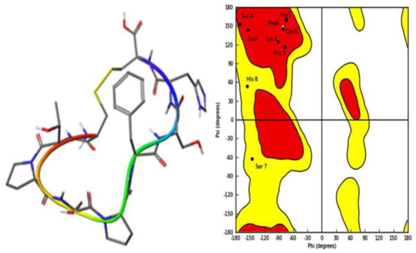
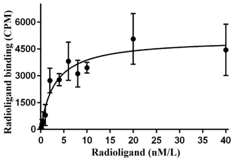
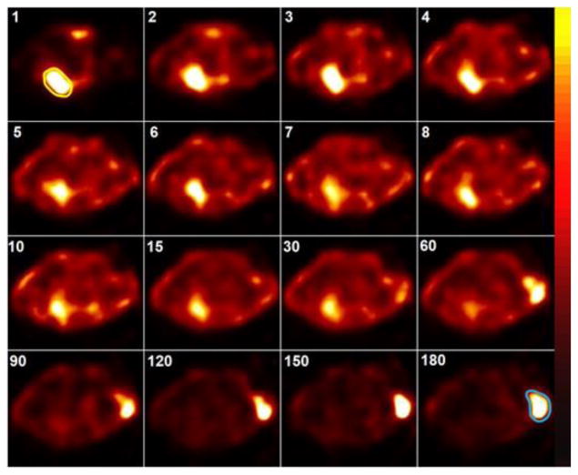
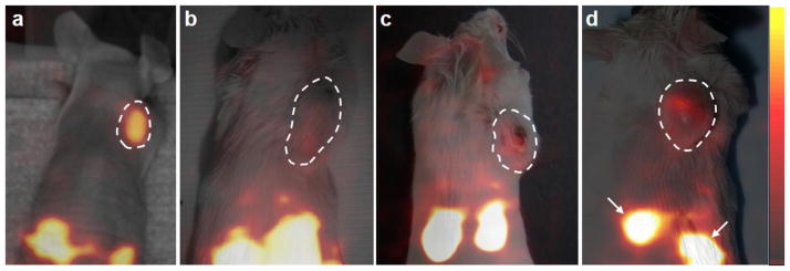
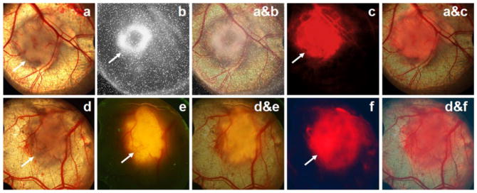
Molecular probes capable of detecting colorectal cancer (CRC) are needed for early CRC diagnosis. The objective of this study was to characterize c[CTPSPFSHC]OH (TCP-1), a small peptide derived from phage display selection, for targeting human CRC xenografts using technetium-99m ((99m)Tc)-labeled TCP-1 and fluorescent cyanine-7 (Cy7)-labeled form of the peptide (Cy7-TCP-1). (99m)Tc-TCP-1 was generated by modifying TCP-1 with succinimidyl-6-hydrazino-nicotinamide (S-HYNIC) followed by radiolabeling. In vitro saturation binding experiments were performed for (99m)Tc-TCP-1 in human HCT116 colon cancer cells. SCID mice with human HCT116 cancer xenografts were imaged with (99m)Tc-TCP-1 or control peptide using a small-animal SPECT imager: Group I (n=5) received no blockade; Group II (n=5) received a blocking dose of non-radiolabeled TCP-1. Group III (n=5) were imaged with (99m)Tc-labeled control peptide (inactive peptide). SCID mice with human PC3 prostate cancer xenografts (Group IV, n=5) were also imaged with (99m)Tc-TCP-1. Eight additional SCID mice bearing HCT116 xenografts in dorsal skinfold window chambers (DSWC) were imaged by direct positron imaging of (18)F-fluorodeoxyglucose ((18)F-FDG) and fluorescence microscopy of Cy7-TCP-1. In vitro(99m)Tc-HYNIC-TCP-1 binding assays on HCT 116 cells indicated a mean Kd of 3.04±0.52nM. In cancer xenografts, (99m)Tc-TCP-1 radioactivity (%ID/g) was 1.01±0.15 in the absence of blockade and was reduced to 0.26±0.04 (P<0.01) with blockade. No radioactive uptake was observed in the PC3 tumors with (99m)Tc-TCP-1 or HCT116 tumors with inactive peptide. Cy7-TCP-1 activity localized not only in metabolically active tumors, as defined by (18)F-FDG imaging, but also in peritumoral microvasculature. In conclusion, TCP-1 probes may have a distinct targeting mechanism with high selectivity for CRC and tumor-associated vasculature. Molecular imaging with TCP-1 probes appears promising to detect malignant colorectal lesions.
Keywords: (99m)Tc; Colorectal cancer; Molecular imaging; Mouse xenograft models; Peptide; SPECT.
Copyright © 2016 Elsevier B.V. All rights reserved.
Figures
Similar articles
-
PEGylated and Non-PEGylated TCP-1 Probes for Imaging of Colorectal Cancer.Mol Imaging Biol. 2023 Feb;25(1):133-143. doi: 10.1007/s11307-021-01684-z. Epub 2021 Nov 29. Mol Imaging Biol. 2023. PMID: 34845659 Free PMC article.
-
[99mTc]Tc-HYNIC-RM2: A potential SPECT probe targeting GRPR expression in prostate cancers.Nucl Med Biol. 2023 Mar-Apr;118-119:108331. doi: 10.1016/j.nucmedbio.2023.108331. Epub 2023 Mar 10. Nucl Med Biol. 2023. PMID: 36933456
-
(99m)Tc-labeled SWL specific peptide for targeting EphA2 receptor.Nucl Med Biol. 2014 Jul;41(6):450-6. doi: 10.1016/j.nucmedbio.2014.03.020. Epub 2014 Mar 29. Nucl Med Biol. 2014. PMID: 24768147
-
In vivo imaging of human colon cancer xenografts in immunodeficient mice using a guanylyl cyclase C--specific ligand.J Nucl Med. 2002 Mar;43(3):392-9. J Nucl Med. 2002. PMID: 11884500
-
Evaluation of (99m)Tc-HYNIC-TMTP1 as a tumor-homing imaging agent targeting metastasis with SPECT.Nucl Med Biol. 2015 Mar;42(3):256-62. doi: 10.1016/j.nucmedbio.2014.11.001. Epub 2014 Nov 13. Nucl Med Biol. 2015. PMID: 25516099
Cited by
-
Monte Carlo study on the secondary cancer risk estimations for patients undergoing prostate radiotherapy: A humanoid phantom study.Rep Pract Oncol Radiother. 2020 Mar-Apr;25(2):187-192. doi: 10.1016/j.rpor.2019.12.029. Epub 2020 Jan 10. Rep Pract Oncol Radiother. 2020. PMID: 32021575 Free PMC article.
-
Identification of a specific peptide binding to colon cancer cells from a phage-displayed peptide library.Br J Cancer. 2018 Jan;118(1):79-87. doi: 10.1038/bjc.2017.366. Epub 2017 Oct 24. Br J Cancer. 2018. PMID: 29065111 Free PMC article.
-
PEGylated and Non-PEGylated TCP-1 Probes for Imaging of Colorectal Cancer.Mol Imaging Biol. 2023 Feb;25(1):133-143. doi: 10.1007/s11307-021-01684-z. Epub 2021 Nov 29. Mol Imaging Biol. 2023. PMID: 34845659 Free PMC article.
-
Novel Peptide-Modified Zeolitic Imidazolate Framework-8 Nanoparticles with pH-Sensitive Release of Doxorubicin for Targeted Treatment of Colorectal Cancer.Pharmaceutics. 2025 Feb 13;17(2):246. doi: 10.3390/pharmaceutics17020246. Pharmaceutics. 2025. PMID: 40006613 Free PMC article.
-
A Dodecapeptide Selected by Phage Display as a Potential Theranostic Probe for Colon Cancers.Transl Oncol. 2020 Sep;13(9):100798. doi: 10.1016/j.tranon.2020.100798. Epub 2020 May 23. Transl Oncol. 2020. PMID: 32454443 Free PMC article.
References
-
- Siegel R, Desantis C, Jemal A. Colorectal cancer statistics, 2014. CA Cancer J Clin. 2014;64:104–117. - PubMed
-
- Triantafillidis JK, Nasioulas G, Kosmidis PA. Colorectal cancer and inflammatory bowel disease: epidemiology, risk factors, mechanisms of carcinogenesis and prevention strategies. Anticancer research. 2009;29:2727–2737. - PubMed
-
- Farraye FA, Odze RD, Eaden J, Itzkowitz SH, McCabe RP, Dassopoulos T, Lewis JD, Ullman TA, James T, 3rd, McLeod R, Burgart LJ, Allen J, Brill JV A.G.A.I.M.P.P.o. Diagnosis, D. Management of Colorectal Neoplasia in Inflammatory Bowel, AGA medical position statement on the diagnosis and management of colorectal neoplasia in inflammatory bowel disease. Gastroenterology. 2010;138:738–745. - PubMed
Publication types
MeSH terms
Substances
Grants and funding
LinkOut - more resources
Full Text Sources
Other Literature Sources
