

Endothelial Dysfunction in Renal Interstitial Fibrosis
- PMID: 27576317
- PMCID: PMC5089917
- DOI: 10.1159/000447607
Endothelial Dysfunction in Renal Interstitial Fibrosis
Abstract
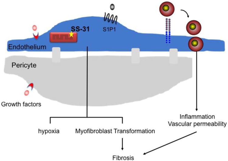
Kidney disease affects millions of people worldwide and it is now widely accepted that many pathological processes may persist after acute kidney injury that can cause the progression to CKD. Tubulointerstitial fibrosis manifests soon after injury and while many cellular and molecular components of kidney fibrosis have been discovered, largely in animal models, new therapeutic strategies are still desperately needed. The renal endothelium has emerged as important in progression of fibrosis through regulation of hypoxia, inflammation and cellular crosstalk. This review aims to highlight our current understanding of the role of the endothelium in interstitial fibrosis and to identify potential therapeutic targets. © 2016 S. Karger AG, Basel.
Figures
References
-
- Sutton TA, Fisher CJ, Molitoris BA. Microvascular endothelial injury and dysfunction during ischemic acute renal failure. Kidney Int. 2002;62:1539–1549. - PubMed
-
- Wu L, Tiwari MM, Messer KJ, Holthoff JH, Gokden N, Brock RW, Mayeux PR. Peritubular capillary dysfunction and renal tubular epithelial cell stress following lipopolysaccharide administration in mice. Am J Physiol Renal Physiol. 2007;292:F261–268. - PubMed
Publication types
MeSH terms
Grants and funding
LinkOut - more resources
Full Text Sources
Other Literature Sources
Medical
