

Nematode infection in liver of the fish Gymnotus inaequilabiatus (Gymnotiformes: Gymnotidae) from the Pantanal Region in Brazil: pathobiology and inflammatory response
- PMID: 27576434
- PMCID: PMC5006381
- DOI: 10.1186/s13071-016-1772-2
Nematode infection in liver of the fish Gymnotus inaequilabiatus (Gymnotiformes: Gymnotidae) from the Pantanal Region in Brazil: pathobiology and inflammatory response
Abstract
Background: A survey on endoparasitic helminths from freshwater fishes in the Pantanal Region (Mato Grosso do Sul, Brazil) revealed the occurrence of third-larval stage of the nematode Brevimulticaecum sp. (Heterocheilidae) in most organs of Gymnotus inaequilabiatus (Gymnotidae) also known by the local name tuvira. The aim of the present study was to examine Brevimulticaecum sp.-infected tuvira liver at the ultrastructural level and clarify the nature of granulomas and the cellular elements involved in the immune response to nematode larvae.
Methods: Thirty-eight adult specimens of tuvira from Porto Morrinho, were acquired in January and March 2016. Infected and uninfected liver tissues were fixed and prepared for histological and ultrastructure investigations.
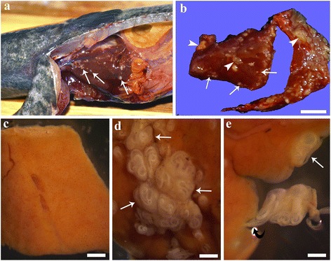
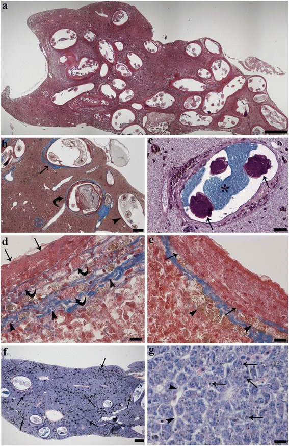
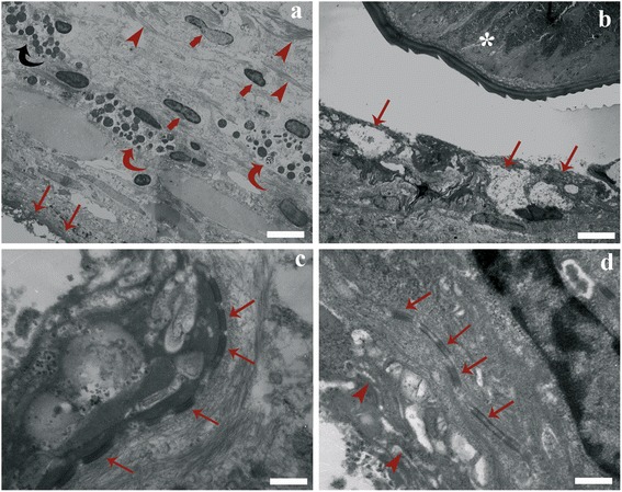
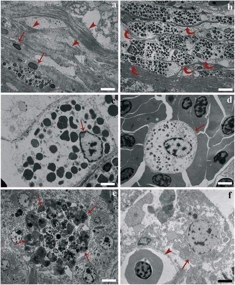
Results: The prevalence of infection of tuvira liver by the nematode larvae was 95 %, with an intensity of infection ranging from 4 to 343 larvae (mean ± SD: 55.31 ± 73.94 larvae per liver). In livers with high numbers of nematode larvae, almost entire hepatic tissue was occupied by the parasites. Hepatocytes showed slight to mild degenerative changes and accumulation of pigments. Parasite larvae were surrounded by round to oval granulomas, the result of focal host tissue response to the infection. Each granuloma was typically formed by three concentric layers: an outer layer of fibrous connective tissue with thin elongated fibroblasts; a middle layer of mast cells entrapped in a thin fibroblast-connective mesh; and an inner layer of densely packed epithelioid cells, displaying numerous desmosomes between each other. Numerous macrophage aggregates occurred in the granulomas and in the parenchyma.
Conclusions: Our results in tuvira showed that the larvae were efficiently sequestered within the granulomas, most of the inflammatory components were confined within the thickness of the granuloma, and the parenchyma was relatively free of immune cells and without fibrosis. Presumably this focal encapsulation of the parasites permits uninfected portions of liver to maintain its functions and allows the survival of the host.
Keywords: Fish immune response; Hepatic granuloma; Histopathology; Nematode larvae.
Figures
Similar articles
-
Liver of the fish Gymnotus inaequilabiatus and nematode larvae infection: Histochemical features and expression of proliferative cell nuclear antigen.J Fish Dis. 2017 Dec;40(12):1765-1774. doi: 10.1111/jfd.12641. Epub 2017 May 11. J Fish Dis. 2017. PMID: 28493503
-
Brevimulticaecum sp. (Nematoda: Heterocheilidae) larvae parasitic in freshwater fish in the Pantanal wetland, Brazil.Vet Parasitol. 2010 Sep 20;172(3-4):350-4. doi: 10.1016/j.vetpar.2010.05.003. Epub 2010 May 11. Vet Parasitol. 2010. PMID: 20684864
-
A portrait of 2 nematodes in liver of their paratenic fish hosts, illustrating different immunological approaches.Parasitology. 2025 Jul 14:1-9. doi: 10.1017/S003118202510053X. Online ahead of print. Parasitology. 2025. PMID: 40653765
-
First record of Trypanosoma sp. (Protozoa: Kinetoplastida) in tuvira (Gymnotus aff. inaequilabiatus) in the Pantanal wetland, Mato Grosso do Sul State, Brazil.Rev Bras Parasitol Vet. 2011 Jan-Mar;20(1):85-7. doi: 10.1590/s1984-29612011000100019. Rev Bras Parasitol Vet. 2011. PMID: 21439241
-
Fish immune responses against endoparasitic nematodes - experimental models.J Fish Dis. 2012 Sep;35(9):623-35. doi: 10.1111/j.1365-2761.2012.01385.x. Epub 2012 Jun 6. J Fish Dis. 2012. PMID: 22671918 Review.
Cited by
-
Pike intestinal reaction to Acanthocephalus lucii (Acanthocephala): immunohistochemical and ultrastructural surveys.Parasit Vectors. 2018 Jul 16;11(1):424. doi: 10.1186/s13071-018-3002-6. Parasit Vectors. 2018. PMID: 30012189 Free PMC article.
-
Stages of Granulomatous Response Against Histozoic Metazoan Parasites in Mullets (Osteichthyes: Mugilidae).Animals (Basel). 2021 May 21;11(6):1501. doi: 10.3390/ani11061501. Animals (Basel). 2021. PMID: 34064270 Free PMC article.
-
Extrusion of Contracaecum osculatum nematode larvae from the liver of cod (Gadus morhua).Parasitol Res. 2017 Oct;116(10):2721-2726. doi: 10.1007/s00436-017-5580-1. Epub 2017 Aug 9. Parasitol Res. 2017. PMID: 28795224
-
The Tilapia Cyst Tissue Enclosing the Proliferating Myxobolus bejeranoi Parasite Exhibits Cornified Structure and Immune Barrier Function.Int J Mol Sci. 2024 May 23;25(11):5683. doi: 10.3390/ijms25115683. Int J Mol Sci. 2024. PMID: 38891869 Free PMC article.
-
Parasites and the neuroendocrine control of fish intestinal function: an ancient struggle between pathogens and host.Parasitology. 2022 Dec;149(14):1842-1861. doi: 10.1017/S0031182022001160. Epub 2022 Aug 16. Parasitology. 2022. PMID: 36076315 Free PMC article. Review.
References
-
- Isaac A, Guidelli GM, Franca JG, Pavanelli GC. Composicao e estrutura das infracomunidades endoparasitarias de Gymnotus spp. (Pisces: Gymnotidae) do rio Baia, Mato Grosso do Sul, Brasil. Acta Scient Biol Sci. 2004;26:453–62.
-
- Catto JB, Amato FFR. Helminth community structure of the Caiman crocodilus yacare in the Brazilian Pantanal. Rev Bras Parasitol Vet. 1994;3:109–18.
-
- Goldberg SR, Bursey CR. Helminths of two species of frogs, Lithobates taylori and Lithobates vaillanti (Ranidae), from Costa Rica. Car J Sci. 2007;43:65–72.
Publication types
MeSH terms
LinkOut - more resources
Full Text Sources
Other Literature Sources
