. 2016 Sep 30;119(8):931-43.
doi: 10.1161/CIRCRESAHA.116.308964. Epub 2016 Aug 30.

Genetically Encoded Biosensors Reveal PKA Hyperphosphorylation on the Myofilaments in Rabbit Heart Failure

Affiliations

Genetically Encoded Biosensors Reveal PKA Hyperphosphorylation on the Myofilaments in Rabbit Heart Failure

Federica Barbagallo et al. Circ Res. .

Abstract

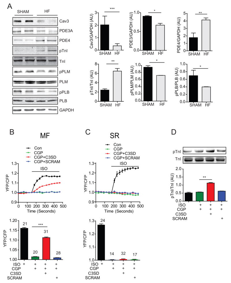
Rationale: In heart failure, myofilament proteins display abnormal phosphorylation, which contributes to contractile dysfunction. The mechanisms underlying the dysregulation of protein phosphorylation on myofilaments is not clear.

Objective: This study aims to understand the mechanisms underlying altered phosphorylation of myofilament proteins in heart failure.

Methods and results: We generate a novel genetically encoded protein kinase A (PKA) biosensor anchored onto the myofilaments in rabbit cardiac myocytes to examine PKA activity at the myofilaments in responses to adrenergic stimulation. We show that PKA activity is shifted from the sarcolemma to the myofilaments in hypertrophic failing rabbit myocytes. In particular, the increased PKA activity on the myofilaments is because of an enhanced β2 adrenergic receptor signal selectively directed to the myofilaments together with a reduced phosphodiesterase activity associated with the myofibrils. Mechanistically, the enhanced PKA activity on the myofilaments is associated with downregulation of caveolin-3 in the hypertrophic failing rabbit myocytes. Reintroduction of caveolin-3 in the failing myocytes is able to normalize the distribution of β2 adrenergic receptor signal by preventing PKA signal access to the myofilaments and to restore contractile response to adrenergic stimulation.

Conclusions: In hypertrophic rabbit myocytes, selectively enhanced β2 adrenergic receptor signaling toward the myofilaments contributes to elevated PKA activity and PKA phosphorylation of myofilament proteins. Reintroduction of caveolin-3 is able to confine β2 adrenergic receptor signaling and restore myocyte contractility in response to β adrenergic stimulation.

Keywords: adrenergic receptor; heart failure; myofibrils; phosphorylation; protein kinase A.


Figures

Figure 1
Figure 1. Generation of cardiac myofilament-targeted FRET biosensors
A) Schematic representation of subcellular-localized PKA AKAR3 biosensors. Yellow (YFP) and cyan (CFP) fluorescent proteins flank a PKA substrate and a forkhead-associated (FHA) domain that recognizes the phosphorylated PKA substrate. AKAR3 is linked to a Kras-derived sequence for plasma membrane (PM-AKAR3) localization, to a phospholamban (PLB)-derived sequence for sarcoplasmic reticulum (SR-AKAR3) localization, and to troponin T for myofilament (MF-AKAR3) localization. B) Representative confocal images of biosensor (green) expressed in young rabbit cardiac myocytes. Cells are immunostained with subcellular specific markers (red) for the SR (Ryanodine Receptor, RyR), the PM (Wheat Germ Agglutinin, WGA), and the MF (Phalloidin). Merge images confirm co-localization of fluorescent signals. C) Immunoblot analyses of PKA phosphorylation of troponin I (TnI) at serine 23/24 in response to isoproterenol (ISO) stimulation (1 or 100 nmol/L) in young rabbit myocytes infected with MF-AKAR3 probes. D) Young rabbit myocytes expressing PM-AKAR3, SR-AKAR3, and MF-AKAR3 are stimulated with a set of incremental doses of ISO. Time courses show AKAR3 FRET responses after stimulation with ISO. E) Normalized isoproterenol-induced dose response curves of AKAR3 biosensors (EC50 PM-AKAR3 at 2.16 × 10⁻¹⁰ mol/L, SR-AKAR3 at 2.57 × 10⁻⁹ mol/L, and MF-AKAR at 3.89 × 10⁻⁹ mol/L). F) Maximal increases in AKAR3 FRET ratio after stimulation with ISO (1 μM) or after co-treatment with forskolin (10 μM) and IBMX (100 μM). G) Normalized maximal FRET responses of individual AKAR3 biosensors against the increases induced by co-treatment with forskolin and IBMX, respectively.
Figure 2
Figure 2. A highly localized β2AR signal is not accessible to the myofilaments in SHAM rabbit cardiac myocytes
A) SHAM rabbit cardiac myocytes expressing MF-AKAR3 are stimulated with 100 nmol/L ISO (total), or in the presence of 100 nmol/L β2AR blocker ICI118551 (β1AR) or 300 nmol/L of the β1AR blocker CGP20712A (β2AR). The maximal increases in MF-AKAR3 are plotted in bar graph. The dash line indicates the maximal increases induced by forskolin (10 μmol/L) and IBMX (100 μmol/L). *** p < 0.01 by one-way ANOVA followed by post hoc Bonferroni test. B) Stimulation of SHAM cardiac myocytes with ISO in the presence of CGP20712A or ICI118551 for 5 min. The PKA phosphorylation of TnI at Ser 23/24 is detected in western blot, and the increases in phosphorylation of TnI are plotted in bar graph. N = 6, ** p < 0.01 by one-way ANOVA followed by post hoc Bonferroni test). C) Time courses of FRET responses are from SHAM and HF cardiac myocytes expressing MF-AKAR3 after stimulation of βARs. D) Bar graph represents the maximal increases in MF-AKAR3 FRET ratio in panel C. * p < 0.05 and *** p < 0.001 by student t-test. E) Stimulation of SHAM and HF cardiac myocytes with ISO in the presence of CGP20712A or ICI118551 for 5 min. The PKA phosphorylation of TnI at Ser 23/24 is detected in western blot and quantified in bar graph. N = 6, ** p < 0.01 by one-way ANOVA followed by post hoc Bonferroni test).
Figure 3
Figure 3. Diminished βAR-induced PKA activity at the SR in failing cardiac myocytes
A) SHAM rabbit cardiac myocytes expressing SR-AKAR3 are stimulated with 100 nmol/L ISO (total), or in the presence of 100 nmol/L β2AR blocker ICI118551 (β1AR) or 300 nmol/L of the β1AR blocker CGP20712A (β2AR). The maximal increases in SR-AKAR3 are plotted in bar graph. The dash line indicates the maximal increases induced by forskolin (10 μmol/L) and IBMX (100 μmol/L). ** p < 0.01 when compared to total by one-way ANOVA followed by post hoc Bonferroni test. B) Stimulation of SHAM cardiac myocytes with ISO in the presence of CGP20712A or ICI118551 for 5 min. The PKA phosphorylation of PLB at serine 16 is detected in western blot, and plotted in bar graph. N = 6, ** p < 0.01 by one-way ANOVA followed by post hoc Bonferroni test). C) Time courses of FRET responses are from SHAM and HF cardiac myocytes expressing SR-AKAR3 after stimulation of βARs. D) Bar graph represents the maximal increases in SR-AKAR3 FRET ratio in panel C. * p < 0.05 by student t-test. E) Stimulation of SHAM and HF cardiac myocytes with ISO in the presence of CGP20712A or ICI118551 for 5 min. The PKA phosphorylation of PLB is detected in western blot and quantified in bar graph. N = 6, ** p < 0.01 and *** p < 0.001 by one-way ANOVA followed by post hoc Bonferroni test.
Figure 4
Figure 4. Redistribution of βAR subtype activity at the PM in failing cardiac myocytes
A) SHAM and HF rabbit cardiac myocytes expressing PM-AKAR3 are stimulated with 100 nmol/L ISO (total), in the presence of 100 nmol/L β2AR blocker ICI118551 (β1AR), or 300 nmol/L of the β1AR blocker CGP20712A (β2AR). Time courses show FRET responses from SHAM and HF cardiac myocytes expressing PM-AKAR3 after stimulation of βARs. B–D) The maximal increases in PM-AKAR3 in SHAM and HF myocytes are plotted in bar graph. In panel B, the dash line indicates the maximal increases induced by forskolin (10 μmol/L) and IBMX (100 μmol/L). # p < 0.05 and ## p < 0.01 by one-way ANOVA followed by post hoc Bonferroni test. * p < 0.05 by student t-test.
Figure 5
Figure 5. Redistribution of PDE activity in failing cardiac myocytes
A–F) SHAM and HF rabbit cardiac myocytes expressing MF-AKAR3, SR-AKAR3, or PM-AKAR3 are treated with 10 μmol/L rolipram (PDE4), or 1 μmol/L cilostamide (PDE3). Time courses of FRET traces from SHAM and HF cardiac myocytes are shown. G–I) The maximal increases in AKAR3 FRET ratio in SHAM and HF myocytes are plotted in bar graph. * p < 0.05 and *** p < 0.001 by student t-test.
Figure 6
Figure 6. Inhibition of caveolin-3 promotes adrenergic signaling on the myofilaments in HF cardiac myocytes
A) Immunoblots show expression of caveolin-3, PDE3A, PDE4, TnI, PLB, and phospholemman (PLM), as well as pTnI at serine 23/24, pPLB at serine 16, and pPLM at serine 68 in SHAM and HF heart lysates. Bar graphs represent mean ± SEM. N = 3; * p < 0.05, ** p < 0.01, and *** p < 0.01 by student’s t-test. B and C) Healthy young rabbit myocytes expressing SR-AKAR3 or MFAKAR3 are stimulated with 100nmol/L ISO and 300 nmol/L CGP20712A after incubation with 1 μmol/L C3SD or scrambled peptide for 1 hour. Time courses of changes in AKAR3 FRET ratio are shown, and the maximal increases in FRET ratio are plotted in bar graph. *** p < 0.001 by one-way ANOVA followed by post hoc Bonferroni test. D) Healthy young rabbit myocytes are stimulated with 100 nmol/L ISO in the presence of 1 μmol/L C3SD or scrambled peptide. PKA phosphorylation of troponin I at serine 23/24 is detected in western blots and quantified in bar graph. N = 3; ** p < 0.01 by one-way ANOVA followed by post hoc Bonferroni test.
Figure 7
Figure 7. Expression of caveolin-3 normalizes adrenergic signaling on the myofilaments in HF cardiac myocytes
A–C) SHAM or HF myocytes expressing MF-AKAR3, PM-AKAR3 or SR-AKAR3 together with caveolin-3 as indicated. Myocytes are stimulated with 100 nmol/L ISO, or in the presence of 100 nmol/L ICI118551 or 300 nmol/L CGP20712A. The maximal increases in FRET ratio are plotted in bar graph. * p < 0.05, ** p < 0.01, and *** p < 0.001 by one-way ANOVA followed by post hoc Bonferroni test. D) HF myocytes expressing caveolin-3 are stimulated with 100 nmol/L ISO in the presence of 300 nmol/L CGP20712A. PKA phosphorylation of troponin I at serine 23/24 are detected in western blots, and quantified in bar graph. N = 5; * p < 0.01 by one-way ANOVA followed by post hoc Bonferroni test. E) SHAM and HF rabbit myocytes with or without expression of caveolin-3 are stimulated with ISO (100 nmol/L). Myocyte contractile shortening is plotted as bar graphs. * p < 0.05 by one-way ANOVA followed by post hoc Bonferroni test.
Figure 8
Figure 8. Schematic models on changes of local PKA signal in failing cardiac myocytes
In SHAM cardiac myocytes, β1AR has a higher and further reaching response in all three subcellular compartments tested than β2AR. β2AR generates a highly localized response within the PM and the SR, but does not induce any significant PKA activity on the myofilaments. PKA activity at the PM microdomain is under predominant control of PDE4 whereas PDE3 preferentially hydrolyzes a pool of cAMP in the vicinity of myofilaments. In HF myocytes with reduced caveolin-3, the βAR-induced PKA activity at the PM and, to a lesser extent, at the SR is reduced. However, the βAR-induced PKA activity at the myofilament is increased due in part to augmented β2AR signal and miss-localization of PDE3 away from the myofilaments. Reintroduction of caveolin-3 normalizes the distribution of βAR-induced PKA activity at the subcellular compartments, including increases in PKA activity at the PM, and decreases in PKA activity at the myofilaments. In particular, the β2AR-induced PKA activity is confined at the PM and the SR, and does not access the myofilaments.

Similar articles

Cited by

References

    1. Ruehr ML, Russell MA, Bond M. A-kinase anchoring protein targeting of protein kinase A in the heart. J Mol Cell Cardiol. 2004;37:653–65. - PubMed
    1. Koss KL, Kranias EG. Phospholamban: a prominent regulator of myocardial contractility. Circ Res. 1996;79:1059–63. - PubMed
    1. Stelzer JE, Patel JR, Walker JW, Moss RL. Differential roles of cardiac myosin-binding protein C and cardiac troponin I in the myofibrillar force responses to protein kinase A phosphorylation. Circ Res. 2007;101:503–11. - PubMed
    1. Eschenhagen T. Beta-adrenergic signaling in heart failure-adapt or die. Nat Med. 2008;14:485–7. - PubMed
    1. Post SR, Hammond HK, Insel PA. Beta-adrenergic receptors and receptor signaling in heart failure. Annu Rev Pharmacol Toxicol. 1999;39:343–60. - PubMed

Substances