

Interleukin-6, MCP-1, IP-10, and MIG are sequentially expressed in cerebrospinal fluid after subarachnoid hemorrhage
- PMID: 27576738
- PMCID: PMC5006407
- DOI: 10.1186/s12974-016-0675-7
Interleukin-6, MCP-1, IP-10, and MIG are sequentially expressed in cerebrospinal fluid after subarachnoid hemorrhage
Abstract
Background: Interleukin-6 (IL-6), an inflammatory cytokine, plays important roles in cerebrospinal fluid (CSF) after subarachnoid hemorrhage (SAH). Chemokines are chemoattractant cytokines that regulate trafficking of monocytes/macrophages and lymphocytes to sites of inflammation. However, no studies have been reported regarding the temporal expression of these cytokines in CSF after SAH.
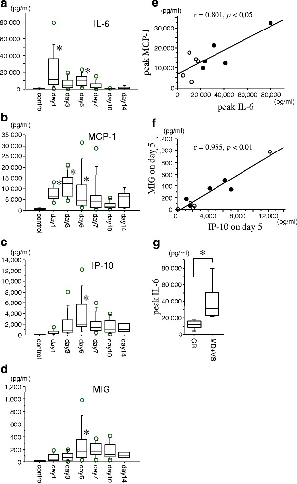
Findings: The concentrations of IL-6, monocyte chemoattractant protein-1 (MCP-1), interferon-γ-inducible protein-10 (IP-10), and monokine induced by interferon-γ (MIG) in the CSF of ten patients with SAH were measured using ELISA kits over a period of 14 days. All aneurysms were located in the anterior circulation. CSF samples from patients with unruptured aneurysms were used as controls. The concentration of IL-6 significantly increased during the acute stage of the disease. The concentration of MCP-1 increased from days 1 to 5, peaking on day 3, and decreased thereafter. The concentrations of IP-10 and MIG progressively increased, peaked on day 5, and then gradually decreased. There were strong correlations between the maximum levels of IL-6 and MCP-1 and IP-10 and MIG on day 5. The maximum level of IL-6 was much higher in poor outcome patients than in good outcome patients.
Conclusions: The present investigation demonstrated that increases in IL-6 levels may induce the expression of MCP-1 in CSF after SAH, followed by increases in the expression of IP-10 and MIG. Dynamic changes in the levels of these cytokines may induce inflammation and may be closely associated with the development of delayed ischemic neurological deficits after SAH.
Keywords: IP-10; Interleukin-6; MCP-1; MIG; Subarachnoid hemorrhage.
Figures
Similar articles
-
Cisternal CSF levels of cytokines after subarachnoid hemorrhage.Neurol Res. 1998 Jun;20(4):337-42. doi: 10.1080/01616412.1998.11740528. Neurol Res. 1998. PMID: 9618698
-
Acute chemokine response in the blood and cerebrospinal fluid of children with enterovirus 71-associated brainstem encephalitis.J Infect Dis. 2008 Oct 1;198(7):1002-6. doi: 10.1086/591462. J Infect Dis. 2008. PMID: 18710325
-
IL-6, IL-8, IP-10, MCP-1 and G-CSF are significantly increased in cerebrospinal fluid but not in sera of patients with central neuropsychiatric lupus erythematosus.Lupus. 2016 Aug;25(9):997-1003. doi: 10.1177/0961203316629556. Epub 2016 Feb 3. Lupus. 2016. PMID: 26846690
-
MCP-1 Is Elevated in the Cerebral Fluid of Children With Tourette Syndrome: Case Series and Literature Review.Brain Behav. 2025 Jun;15(6):e70617. doi: 10.1002/brb3.70617. Brain Behav. 2025. PMID: 40495462 Free PMC article. Review.
-
Evaluation of Chemokines MIG and IP-10 as Immunological Biomarkers of Human Visceral Leishmaniasis: A Systematic Review.Trop Med Infect Dis. 2024 Sep 19;9(9):219. doi: 10.3390/tropicalmed9090219. Trop Med Infect Dis. 2024. PMID: 39330908 Free PMC article. Review.
Cited by
-
The Emerging Role of Pericyte-Derived Extracellular Vesicles in Vascular and Neurological Health.Cells. 2022 Oct 2;11(19):3108. doi: 10.3390/cells11193108. Cells. 2022. PMID: 36231071 Free PMC article. Review.
-
Interleukin-6 in Cerebrospinal Fluid Small Extracellular Vesicles as a Potential Biomarker for Prognosis of Aneurysmal Subarachnoid Haemorrhage.Neuropsychiatr Dis Treat. 2021 May 11;17:1423-1431. doi: 10.2147/NDT.S304394. eCollection 2021. Neuropsychiatr Dis Treat. 2021. PMID: 34012263 Free PMC article.
-
Expression of MMP-9 and IL-6 in patients with subarachnoid hemorrhage and the clinical significance.Exp Ther Med. 2018 Feb;15(2):1510-1514. doi: 10.3892/etm.2017.5553. Epub 2017 Nov 23. Exp Ther Med. 2018. PMID: 29434735 Free PMC article.
-
Dynamics of cytokine and antibody responses in community versus hospital SARS-CoV-2 infections.Front Immunol. 2024 Nov 22;15:1468871. doi: 10.3389/fimmu.2024.1468871. eCollection 2024. Front Immunol. 2024. PMID: 39650666 Free PMC article.
-
Interleukin-6: Important Mediator of Vasospasm Following Subarachnoid Hemorrhage.Curr Neurovasc Res. 2021;18(3):364-369. doi: 10.2174/1567202618666211104122408. Curr Neurovasc Res. 2021. PMID: 34736380 Free PMC article. Review.
References
-
- Nagata K, Sasaki T, Mori T, Nikaido H, Kobayashi E, Kim P, Kirino T. Cisternal talc injection in dog can induce delayed and prolonged arterial constriction resembling cerebral vasospasm morphologically and pharmacologically. Surg Neurol. 1996;45:442–447. doi: 10.1016/0090-3019(95)00455-6. - DOI - PubMed
Publication types
MeSH terms
Substances
LinkOut - more resources
Full Text Sources
Other Literature Sources
Research Materials
Miscellaneous
