Review
. 2016 Nov;25(11):1965-1976.
doi: 10.1002/pro.3030. Epub 2016 Sep 16.

Structural aspects of HDAC8 mechanism and dysfunction in Cornelia de Lange syndrome spectrum disorders

Affiliations
Review

Structural aspects of HDAC8 mechanism and dysfunction in Cornelia de Lange syndrome spectrum disorders

Matthew A Deardorff et al. Protein Sci. 2016 Nov.

Abstract

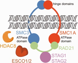
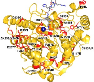
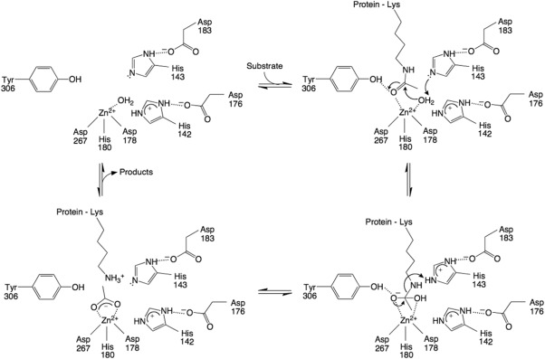
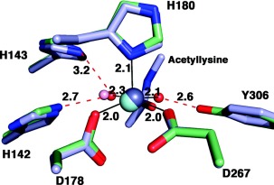
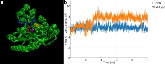
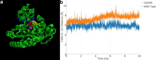
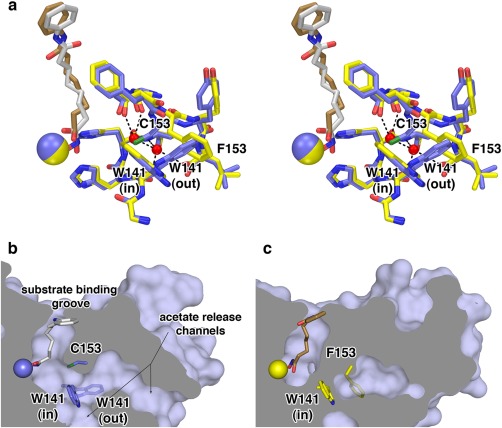
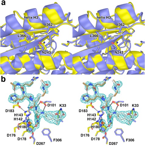
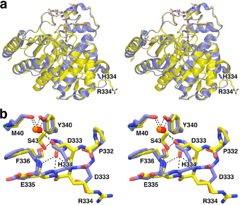
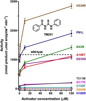
Cornelia de Lange Syndrome (CdLS) encompasses a broad spectrum of phenotypes characterized by distinctive craniofacial abnormalities, limb malformations, growth retardation, and intellectual disability. CdLS spectrum disorders are referred to as cohesinopathies, with ∼70% of patients having a mutation in a gene encoding a core cohesin protein (SMC1A, SMC3, or RAD21) or a cohesin regulatory protein (NIPBL or HDAC8). Notably, the regulatory function of HDAC8 in cohesin biology has only recently been discovered. This Zn2+ -dependent hydrolase catalyzes the deacetylation of SMC3, a necessary step for cohesin recycling during the cell cycle. To date, 23 different missense mutants in the gene encoding HDAC8 have been identified in children with developmental features that overlap those of CdLS. Enzymological, biophysical, and structural studies of CdLS HDAC8 protein mutants have yielded critical insight on compromised catalysis in vitro. Most CdLS HDAC8 mutations trigger structural changes that directly or indirectly impact substrate binding and catalysis. Additionally, several mutations significantly compromise protein thermostability. Intriguingly, catalytic activity in many HDAC8 mutants can be partially or fully restored by an N-acylthiourea activator, suggesting a plausible strategy for the chemical rescue of compromised HDAC8 catalysis in vivo.

Keywords: birth defect; human genetics; lysine deacetylase; protein crystallography; zinc enzyme.


Figures

Figure 1
Figure 1
Cohesin. The cohesin complex consists of four main protein components that encircle sister chromatids during cell division: Structural Maintenance of Chromosomes proteins SMC1A and SMC3, Radiation‐Sensitive Mutant 21 protein RAD21, and Stromal Antigen protein(s) STAG1 or STAG2. ESCO1 and ESCO2 catalyze the acetylation of tandem lysine residues K105 and K106 in SMC3, and HDAC8 catalyzes the deacetylation of these lysine residues to ensure efficient cohesin recycling.
Figure 2
Figure 2
CdLS HDAC8 mutations. A total of 23 different missense mutants have been identified to date in children diagnosed with Cornelia de Lange Syndrome spectrum disorders. Mutations (red) are mapped onto the crystal structure of the Y306F HDAC8‐substrate complex (PDB 2V5W). The active site Zn2+ ion is a dark blue sphere, and 2 K+ ions that regulate HDAC8 activity are orange spheres. The bound substrate is a stick‐figure color‐coded as follows: C = gray, N = blue, O = red.
Figure 3
Figure 3
Proposed mechanism of HDAC8. The Zn2+ ion and Y306 orient and activate the substrate carbonyl for nucleophilic attack by a Zn2+‐bound water molecule, assisted by general base H143. The tetrahedral intermediate and its flanking transition states are stabilized by Zn2+ and hydrogen bond interactions with H142, H143, and Y306; H142 remains protonated throughout catalysis and serves as an electrostatic catalyst. H143 subsequently serves as a general acid catalyst to protonate the leaving amino group to enable the collapse of the tetrahedral intermediate. Reprinted with permission from ref. 40. Copyright 2016, American Chemical Society.
Figure 4
Figure 4
Zn2+ coordination polyhedron in HDAC8. The active site Zn2+ ion of HDAC8 is coordinated with square pyramidal geometry: D178, D267, and two solvent molecules occupy equatorial positions, and H180 occupies the apical position, as observed in the unliganded active site of D101L HDAC8 (PDB 3EW8). Atoms are color coded as follows: C= light green, N = blue, O = red, Zn2+ = gray sphere, water molecules = red spheres. Metal coordination and hydrogen bond interactions are shown as solid black and dotted red lines, respectively. Coordinates of the Y306F HDAC8‐substrate complex are superimposed (PDB 2V5W), with atoms color coded similarly except that C = light blue, Zn2+ = cyan sphere, and water = pink sphere. Note that the substrate carbonyl displaces one of the Zn2+‐bound solvent molecules and accepts a hydrogen bond from Y306. Reprinted with permission from ref 36. Copyright 2008, American Chemical Society.
Figure 5
Figure 5
H180R HDAC8. (a) MD simulation of H180R HDAC8 (superimposed 1‐ns snapshots from a 10‐ns simulation). For reference, a tetrapeptide substrate (blue) as bound to H143A HDAC8 (PDB 3EWF) is superimposed to indicate the location of the active site. Zn2+ is a magenta sphere, R180 is red, and Y306 is yellow. The R180 side chain partially overlaps with the scissile acetyllysine side chain of the substrate, indicating that the mutant side chain sterically blocks substrate binding. (b) In a 10‐ns MD simulation of H180R HDAC8, Y306 fluctuates away from the “in” conformation required for catalysis; in a 10‐ns simulation of wild‐type HDAC8, Y306 remains in its catalytically competent conformation. Reprinted with permission from ref. 27. Copyright 2015, American Chemical Society.
Figure 6
Figure 6
G304R HDAC8. (a) MD simulation of G304R HDAC8 (superimposed 1‐ns snapshots from a 10‐ns simulation). For reference, a tetrapeptide substrate (blue) as bound to H143A HDAC8 (PDB 3EWF) is superimposed to indicate the location of the active site. Zn2+ is a magenta sphere, R180 is red, and Y306 is yellow. The R304 side chain partially overlaps with the scissile acetyllysine side chain of the substrate, indicating that the mutant side chain sterically blocks substrate binding. (b) In a 10‐ns MD simulation of G304R HDAC8, Y306 fluctuates away from the “in” conformation required for catalysis; in a 10‐ns simulation of wild‐type HDAC8, Y306 remains in its catalytically competent conformation. Reprinted with permission from ref. 27. Copyright 2015, American Chemical Society.
Figure 7
Figure 7
C153F HDAC8. (a) Superimposition of the HDAC8 C153F‐SAHA complex (C = yellow, N = blue, O = red, S = green, Zn2+ = yellow sphere, SAHA = tan) and the wild‐type HDAC8‐SAHA complex (PDB 1T69) (C = blue, Zn2+ = blue sphere, SAHA = gray). Water molecules (red spheres) occupy the space previously occupied by the C153 side chain. The F153 side chain sterically locks W141 in the “in” conformation. (b) The solvent‐accessible surface of wild‐type HDAC8 shows that the W141 side chain, which is in equilibrium between the “in” and “out” conformations, serves as a gate for acetate release channels. When W141 is in the “out” conformation, the channels are open. (c) Corresponding view of C153F HDAC8 shows that when W141 is locked in the “in” conformation by the F153 side chain, the acetate release channels are completely blocked. Reprinted with permission from ref. 26. Copyright 2014, American Chemical Society.
Figure 8
Figure 8
I243N HDAC8. (a) Comparison of the I243N HDAC8‐SAHA complex (yellow, C = yellow, N = blue, O = red) with the wild‐type HDAC8‐SAHA complex (blue; PDB 1T69). Selected residues are indicated. Helix H3 shifts by 0.3–1.6 Å as a result of the mutation. (b) Comparison of the I243N/Y306F HDAC8‐substrate complex (C = yellow (protein) or tan (substrate), N = blue, O = red, Zn2+ = yellow sphere) and the Y306F HDAC8‐substrate complex (C = blue (protein) or gray (substrate), N = blue, O = red, Zn2+ = blue sphere) (PDB 2V5W). Water molecules are indicated as red or orange spheres, respectively. Metal coordination and hydrogen bond interactions are shown as solid black and dashed black lines, respectively. The simulated annealing omit map is contoured at 3.0σ and shows a nearly fully ordered tetrapeptide substrate bound in the active site of I243N/Y306F HDAC8. Reprinted with permission from ref. 26. Copyright 2014, American Chemical Society.
Figure 9
Figure 9
H334R HDAC8. (a) Superposition of the H334R/Y306F HDAC8‐substrate complex (yellow) and the Y306F HDAC8‐substrate complex (blue; PDB 2V5W). (b) Close‐up view of local structural changes resulting from the H334R substitution; hydrogen bond interactions are shown as dashed black lines. Atomic color codes are as follows: C = yellow, N = blue, O = red, S = green, water molecule = red sphere. Reprinted with permission from ref. 26. Copyright 2014, American Chemical Society.
Figure 10
Figure 10
Rescue of catalysis in CdLS HDAC8 mutants by TM251. The activity level for wild‐type HDAC8 in the absence of activator is indicated by a dashed line. The catalytic activities of several mutants can be restored to wild‐type levels in dose‐dependent fashion by TM251. All but the catalytically dead mutants H180R and G304R exhibit at least some tendency for activation. Reprinted with permission from ref. 27. Copyright 2015, American Chemical Society.

Similar articles

Cited by

References

    1. Liu J, Krantz ID (2009) Cornelia de Lange syndrome, cohesin, and beyond. Clin Genet 76:303–314. - PMC - PubMed
    1. Dorsett D, Krantz ID (2009) On the molecular etiology of Cornelia de Lange syndrome. Ann NY Acad Sci 1151:22–37. - PMC - PubMed
    1. Liu J, Baynam G (2010) Cornelia de Lange syndrome. Adv Exp Med Biol 685:111–123. - PubMed
    1. Boyle MI, Jespersgaard C, Brøndum‐Nielsen K, Bisgaard A‐M, Tümer Z (2015) Cornelia de Lange syndrome. Clin Genet 88:1–12. - PubMed
    1. Barisic I, Tokic V, Loane M, Bianchi F, Calzolari E, Garne E, Wellesley D, Dolk H, EUROCAT Working Group (2008) Descriptive epidemiology of Cornelia de Lange syndrome in Europe. Am J Med Genet a 146A:51–59. - PubMed

Publication types

MeSH terms

LinkOut - more resources