. 2016 Aug 31:6:32062.
doi: 10.1038/srep32062.

Doxorubicin Conjugated to Immunomodulatory Anticancer Lactoferrin Displays Improved Cytotoxicity Overcoming Prostate Cancer Chemo resistance and Inhibits Tumour Development in TRAMP Mice

Affiliations

Doxorubicin Conjugated to Immunomodulatory Anticancer Lactoferrin Displays Improved Cytotoxicity Overcoming Prostate Cancer Chemo resistance and Inhibits Tumour Development in TRAMP Mice

Jayanth Suryanarayanan Shankaranarayanan et al. Sci Rep. .

Erratum in

Abstract

Advanced, metastatic, castration resistant and chemo-resistant prostate cancer has triggered change in the drug development landscape against prostate cancer. Bovine lactoferrin (bLf) is currently attracting attention in clinics for its anti-cancer properties and proven safety profile. bLf internalises into cancer cells via receptor mediated endocytosis, boosts immunity and complements chemotherapy. We employed bLf as an excellent functional carrier protein for delivering doxorubicin (Dox) into DU145 cells, CD44+/EpCAM+ double positive enriched DU145 3D prostaspheres and drug resistant ADR1000-DU145 cells, thus circumventing Dox efflux, to overcome chemo-resistance. Successful bLf-Dox conjugation with iron free or iron saturated bLf forms did not affect the integrity and functionality of bLf and Dox. bLf-Dox internalised into DU145 cells within 6 h, enhanced nuclear Dox retention up to 24 h, and proved significantly effective (p < 0.001) in reducing LC50 value of Dox from 5.3 μM to 1.3 μM (4 fold). Orally fed iron saturated bLf-Dox inhibited tumour development, prolonged survival, reduced Dox induced general toxicity, cardiotoxicity, neurotoxicity in TRAMP mice and upregulated serum levels of anti-cancer molecules TNF-α, IFN-γ, CCL4 and CCL17. The study identifies promising potential of a novel and safer bLf-Dox conjugate containing a conventional cytotoxic drug along with bLf protein to target drug resistance.


Figures

Figure 1
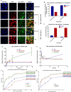
Figure 1. Synthesis and characterisation of bLf-Dox conjugates.
(A) Image indicates the purity of synthesised forms of bLf-Dox conjugates prepared from Apo-bLf and Fe-bLf using SDS PAGE represented by a single non-denatured band around 78 kDa. (B) Western blot was carried out for the synthesised bLf-Dox conjugates against goat anti-bLf monoclonal antibody (1:1000) showing antigen reactivity ~78 kDa. (C) FTIR spectroscopy analysis was performed between 4000 and 400 cm−1 at a resolution of 4 cm−1 averaging 10 scans. The spectrum of each sample was then plotted with percentage transmittance against wavenumber. Crucial peaks are highlighted in the spectra. (D) The thermal stability and the crystallinity of the conjugates was studied using differential scanning calorimetry (DSC) and the endotherm has been represented. (E) The secondary structural characteristics of Apo-bLf and Fe-bLf before and after Dox conjugation was studied using Circular Dichroism (CD) spectroscopy. CD values were obtained for secondary structure prediction at a wavelength range of 250 nm to 190 nm at an interval rate of 0.1 nm/s and the resultant CD spectra of milli degrees vs wavelength was plotted in the graph. The functional stability of Dox in the form of conjugates was confirmed by its ability to inhibit human topoisomerase II (TOPO II) to decatenated human kinetochore DNA (kDNA). The presence of nicked circular kDNA and relaxed circular kDNA was considered as a positive for the activity of TOPO II uninhibited by Dox. The presence of only the catenated kDNA at the wells is considered complete inhibition of TOPO II by Dox.
Figure 2
Figure 2. bLf-Dox conjugates promote greater retention of Dox resulting in enhanced cancer cytotoxicity.
(A) Representative confocal microscopy images of DU145 cells showing the time dependent internalisation of Apo-bLf-Dox and Fe-bLf-Dox studied by immunofluorescence using goat anti-bLf primary antibody (1:100) and anti-goat IgG FITC conjugated secondary antibody (1:100). The internalisation of Dox was studied using its auto-fluorescence with excitation at 488 nm and emission at 630 nm. The nucleus was counterstained with DAPI indicated by the blue fluorescence along with a competitive binding to Dox. Scale = 25 μm. (B) The percentage of cells showing green and red fluorescence co-localised in cytoplasm (yellow) was considered as percentage bLf conjugated Dox plotted as histogram mean ± S.D. (C) Histogram showing presence of nuclear red fluorescence within the cells among 100 counted cells (mean ± S.D). (D) The amount of Dox present within the cells at various time points was considered as Dox retention and the relative fluorescence was plotted against time. Measurements were performed thrice in triplicates and the results are represented as mean ± S.D. (E) The amount of Dox present in the supernatant of the cells treated at various time points was considered as Dox retention and the relative fluorescence was plotted against time. Measurements were performed thrice in triplicates and the results are represented as mean ± S.D. (F) Cytotoxicity determination based on the LDH release from the cells post 24 h treatment at different concentrations in DU145 cells. The experiment was carried out thrice in triplicates. The X-axis is represented in terms of (F). bLf protein concentrations and (G). Dox concentrations. The determination of LC50 was carried out by non-linear regression fitted curve of the cytotoxicity against concentration and values are represented in the graph.
Figure 3
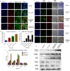
Figure 3. bLf-Dox conjugates treatment induces cancer cell apoptosis.
(A) Representative confocal microscopy images of DU145 cells showing the expression of annexin-V as a marker of apoptosis post treatment with Dox, Apo-bLf-Dox and Fe-bLf-Dox. The annexin-V was stained using immunofluorescence with rabbit anti-annexin-V primary antibody and anti-rabbit-IgG FITC conjugated secondary antibody represented in green fluorescence. Dox auto-fluorescence is represented in red. The nucleus was counterstained with DAPI (blue). Scale bar = 50 μm. (B) Percentage of cellular annexin-V expression from 5 images each was calculated and represented as a histogram (Mean ± S.D.). (C) The presence of fragmented DNA as an end product of apoptotic cascade within the cell was considered as the confirmatory test for induction of apoptosis especially the Dox mediated DNA damage using TUNEL assay. The nucleus was counterstained with DAPI and Dox auto-fluorescence is represented in red. (D) Percentage of TUNEL positive cells from 5 images each has been represented as a histogram. Statistical analysis was performed using one-way ANOVA and a post-hoc Tukey’s test. (E) The molecular regulation of apoptosis was studied using Western blots for various apoptotic markers. The Western blotting was carried out with 100 μg of complete cell lysates against respective primary and secondary antibodies. (F) The Western blot images acquired were analysed for band density using ImageJ (NIH) software and the protein expression was given in relative fold change as compared to the untreated sample normalised against GAPDH and expressed as a histogram.
Figure 4
Figure 4. bLf-Dox conjugates are capable of overcoming molecular drug resistance pathway.
(A) Representative confocal microscopy images of DU145 cells showing the expression of P-gp as a marker of drug resistance. The P-gp was stained using immunofluorescence with mouse anti-P-gp primary antibody and anti-mouse-IgG FITC secondary antibody visualised in green fluorescence. Dox–Red; Nucleus–Blue. Reduced DAPI fluorescence was observed in treatment involving Dox due to competitive nuclear binding. (B) Percentage of cellular P-gp expression was calculated as an average from 5 images for which data has been represented as a histogram (B). Statistical analysis was performed using multiple Student’s t-tests. (C) The molecular regulation of drug resistance was studied using Western blots for various drug resistance markers. The Western blotting was carried out with 100 μg of complete cell lysates against respective primary and secondary antibodies. (D) The Western blot images acquired were analysed for band density using Image J (NIH) software and the protein expression was given in relative fold change as compared to the untreated sample normalised against GAPDH and expressed as a histogram.
Figure 5
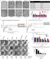
Figure 5. Advanced drug resistant (ADR1000-DU145) cells were sensitive to bLf-Dox treatments but not Dox alone.
(A) The images represent the morphological changes in the DU145 cells upon subjected to pulse exposure of increasing concentration of Dox from 1 nM to 1000 nM. The cells accustomed to 640 nM Dox were treated with 1000 nM Dox on alternate days in complete growth media. The Dox treated DU145 cells were photographed every day following the first treatment and the different stages the cells undergo before they become accustomed to 1000 nM Dox has been represented. These cells were considered Advanced Dox Resistant at 1000 nM (ADR1000-DU145 cells). (B) Doubling time of cells subjected to development of chemo-resistance with Dox. (C) qRT-PCR was performed on the cDNA of DU145 cells and ADR 1000-DU145 cells. The relative fold expression changes of 9 genes of drug resistance and CSC markers were studied and is represented as bar graph (Livak Method). All values are represented as mean ± S.D and the experiment was done twice in duplicates. (D) Cytotoxicity of bLf-Dox conjugates on ADR1000-DU145 cells as a measure of LDH release from the cells was calculated after 24 h treatment at different concentrations. The LC50 values are represented mean ± S.D against respective Dox concentration. (E) Cytotoxicity (LC50) of bLf-Dox conjugates on ADR1000-DU145 cells represented as a function of protein concentrations. (F) ADR1000-DU145 cells were allowed to form prostaspheres for 7 days. Following spheroid formation, they were treated once at 0 h and again at 48 h and the spheroids were imaged under the microscope. (G) The size (Diameter) of the tumour spheroid was measured at 24 h, 48 h and 96 h post first treatment was measured using Image J (NIH) software tool. The experiment was carried out thrice with 5 spheroids per treatment. Two-way ANOVA with post-hoc Tukey’s test was used for statistical analysis. (H) Trypan blue dye exclusion analysis was performed post 96 h treatment to analyse the percentage viable cells left in the spheroids.
Figure 6
Figure 6. Fe-bLf-Dox conjugates inhibited tumour development and prolonged the survival of TRAMP mice.
(A) Kaplan—Meier survival analysis of TRMP mice groups (Control, Dox IP and Fe-bLf-Dox diets) for n = 9 mice per group following treatment post 18 weeks of age for 45 days. (B) Dissected Urino-Genital Tract (UGT) of the TRAMP mice displaying seminal vesicles (S.V), bladder (B), testes (T) and the prostate (Red = Tumourous; Green = Normal). (C) Normalised UGT weights against the mice body weights as a measure of tumour development in TRAMP mice in control, Dox IP and Fe-bLf-Dox feed groups (n = 9). Two-way ANOVA was performed to evaluate statistical significance followed by post-hoc analysis by Tukey’s test. (D) Histopathological analysis of TRAMP mice tissues post haematoxylin and eosin staining viewed under optica light microscope 400X magnifications. Sections of mice intestine, heart, spleen and prostate are provided. (E) Proteome profiler array analysis of serum cytokine expression profile of TRAMP mice treated with Dox and Fe-bLf-Dox (n = 3). Image J analysis of micro-array profile was carried out and relative fold change in the integrated density of each cytokine has been represented as histogram (Mean ± S.D.).
Figure 7
Figure 7. Schematic representation summarizing the inferences of this study about the role of Fe-bLf-Dox conjugates in overcoming drug resistance.
Dox alone (Left) causes increase in chemo-resistance by up-regulating P-gp and MRP-1 expression thereby getting effluxed out of the cancer cells quickly causing minimal damage. bLf-Dox conjugates are easily internalised by cancer cells without triggering P-gp increase and facilitates longer nuclear retention of Dox increasing its cytotoxicity. Meanwhile bLf also decreases survivin, P-gp and MRP-1 expression and increases PTEN expression to decrease chemo-resistance and trigger cancer cell apoptosis. Fe-bLf-Dox treatment in TRAMP mice also reversed the RBC and HGB loss due to Dox treatments and induced anti-tumour immunogenicity. The schematic illustration was generated by modifying images purchased in the PPT Drawing Toolkits-BIOLOGY Bundle from Motifolio, Inc.

Similar articles

Cited by

References

    1. Yagoda A. & Petrylak D. Cytotoxic chemotherapy for advanced hormone‐resistant prostate cancer. Cancer 71, 1098–1109 (1993). - PubMed
    1. Tewey K., Rowe T., Yang L., Halligan B. & Liu L. Adriamycin-induced DNA damage mediated by mammalian DNA topoisomerase II. Science 226, 466–468 (1984). - PubMed
    1. Torti F. M. et al.. Weekly doxorubicin in endocrine-refractory carcinoma of the prostate. Journal of Clinical Oncology 1, 477–482 (1983). - PubMed
    1. Sella A. et al.. Phase II study of ketoconazole combined with weekly doxorubicin in patients with androgen-independent prostate cancer. Journal of Clinical Oncology 12, 683–688 (1994). - PubMed
    1. Collins L., Zhu T., Guo J., Xiao Z. & Chen C. Phellinus linteus sensitises apoptosis induced by doxorubicin in prostate cancer. British Journal of Cancer 95, 282–288 (2006). - PMC - PubMed

Publication types

MeSH terms