. 2016 Sep 13;7(37):58759-58767.
doi: 10.18632/oncotarget.11643.

Efficacy of ATR inhibitors as single agents in Ewing sarcoma

Affiliations

Efficacy of ATR inhibitors as single agents in Ewing sarcoma

Maria Nieto-Soler et al. Oncotarget. .

Abstract

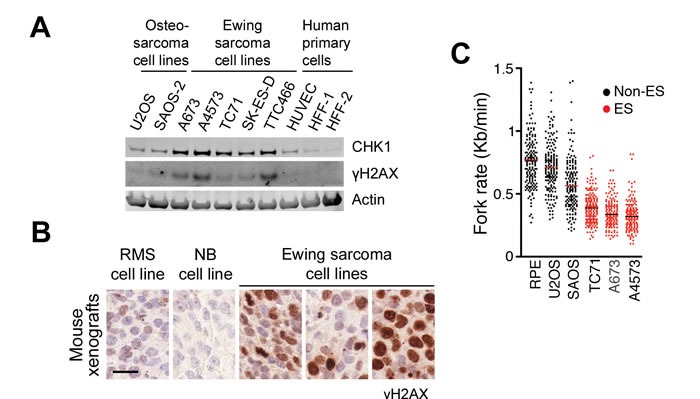
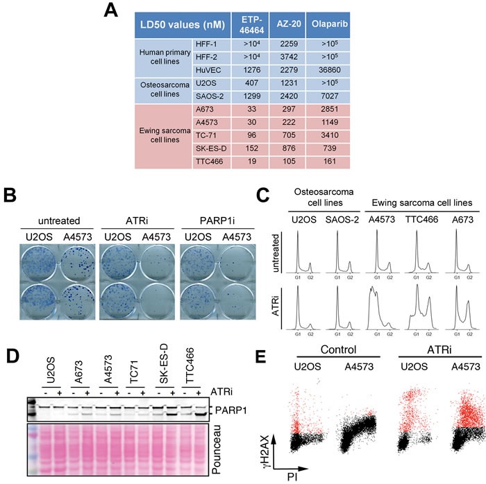
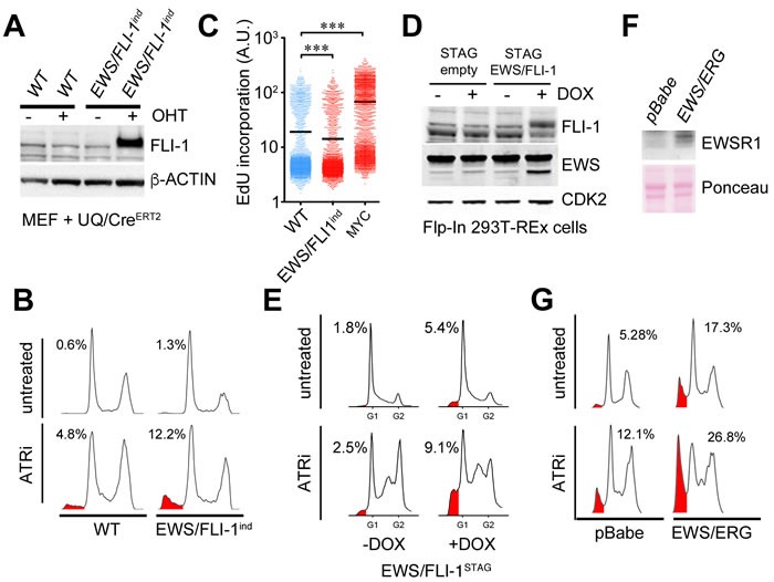
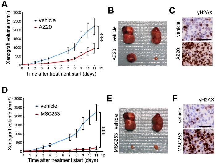
Ewing sarcomas (ES) are pediatric bone tumors that arise from a driver translocation, most frequently EWS/FLI1. Current ES treatment involves DNA damaging agents, yet the basis for the sensitivity to these therapies remains unknown. Oncogene-induced replication stress (RS) is a known source of endogenous DNA damage in cancer, which is suppressed by ATR and CHK1 kinases. We here show that ES suffer from high endogenous levels of RS, rendering them particularly dependent on the ATR pathway. Accordingly, two independent ATR inhibitors show in vitro toxicity in ES cell lines as well as in vivo efficacy in ES xenografts as single agents. Expression of EWS/FLI1 or EWS/ERG oncogenic translocations sensitizes non-ES cells to ATR inhibitors. Our data shed light onto the sensitivity of ES to genotoxic agents, and identify ATR inhibitors as a potential therapy for Ewing Sarcomas.

Keywords: ATR; DNA repair; Ewing sarcoma; cancer; replication stress.


Conflict of interest statement

CONFLICTS OF INTEREST

O.F. receives consulting fees from Merck KGaA. The rest of the authors have no competing financial interests to disclose.

Figures

Figure 1
Figure 1. Increased RS levels in Ewing sarcomas
A. CHK1 and γH2AX levels evaluated by WB on several ES lines, together with 2 osteosarcoma lines and 3 human primary cell types. B. γH2AX IHC on mouse xenografts from 3 ES lines (A4573, A673 and TC71), and two independent xenografts from ES-related tumors (rhabdomyosarcoma (RMS); neuroblastoma (NB)). Scale bar (black) indicates 20 μm. C. Fork rates were measured in stretched DNA fibers prepared from non-ES (RPE, U2OS, SAOS) and ES (TC71, A673 and A4573) cell lines. At least 200 tracks were measured per condition. ***P<0.001 by two-tailed t test.
Figure 2
Figure 2. Sensitivity of ES to ATR inhibitors in vitro
A. LD50 values of 2 independent inhibitors (ETP-46464 [26] and AZ20 [27]) and a PARP1 inhibitor (olaparib, PARPi hereafter) on the same lines used in Figure 1B. The LD50 values for temozolomide, currently used in ES chemotherapy, were above 100 μM in all lines tested. B. Clonogenic assays illustrating the differential effects of ATRi and PARPi on U2OS and A4573 cells. C. DNA content was assessed by flow cytometry on 2 non-ES osteosarcoma lines and 3 ES lines exposed to ATRi for 72 hrs (1 μm). D. Western blot illustrating the cleavage of PARP1 on ES lines and U2OS upon a short exposure to ATRi (1 μM, 4 hrs). E. FACS analysis of DNA content (PI) and H2AX phosphorylation in U2OS and A4573 cells exposed to ATRi (10 μM, 5 hrs), illustrating the increased levels of ATRi-induced RS (as measured by γH2AX in cells with an S-phase DNA content) in ES cells.
Figure 3
Figure 3. Expression of EWSR1 translocations sensitizes cells to ATRi
A. WB illustrating the expression of EWS/FLI1 (measured with a FLI1 antibody) that can be obtained in EWS/FLI1ind MEF upon 4-hydroxy-tamoxifen (OHT)-induced activation of a Cre-ERT2 expressed from the ubiquitin promoter (UQ/CreERT2) [41]. OHT was added for 48 hrs at 1 μM. β-ACTIN was used as loading control. B. DNA content analyses by flow cytometry illustrating the toxicity of ATRi (5μM, 48 hrs) on WT and EWS/FLI1ind MEF harboring UQ-CreERT2 exposed to OHT (1 μM, 48 hrs). SubG1 populations are shaded in red and their percentages are indicated. C. DNA replication rates of WT and EWS/FLI1ind MEF harboring UQ-CreERT2 exposed to OHT, as well as of WT MEF infected with a retrovirus expressing the MYC oncogene were evaluated by quantifying the incorporation of EdU per nucleus by High Throghput Microscopy. D. WB illustrating the expression of EWS/FLI1 (measured with EWS and FLI-1 antibodies) that can be obtained in Flip-In 293T-Rex cells carrying a STAG-EWS/FLI1 cDNA (EWS/FLI1STAG) upon induction with doxycycline (Dox) (200 ng/ml, 48 hrs). The levels in a clone of Flip-In 293T-Rex cells expressing only the STAG peptide are shown as expression controls. CDK2 was used as loading control. E. DNA content analyses by flow cytometry illustrating the toxicity of ATRi (1μM, 24 hrs) on EWS/FLI1STAG cells exposed or not to Dox (48 hrs). SubG1 populations are shaded in red and their percentages are indicated. F. WB illustrating the expression of EWS/ERG (measured with an EWSR1 antibody) that can be obtained in MEF upon infection with a EWS/ERG expressing retrovirus (or empty vector; pBabe). β-ACTIN was used as loading control. G. Flow cytometry illustrating the toxicity of ATRi (5μM, 48 hrs) on MEF infected with an EWS/ERG expressing retrovirus (or empty vector). SubG1 populations are shaded in red and their percentages are indicated.
Figure 4
Figure 4. Efficacy of ATR inhibitors in ES xenografts as single agents
A. Efficacy of AZ20 as monotherapy on the growth of ES xenografts (A4573). Treatment started when tumors became palpable. B. Examples of the tumor sizes observed at endpoint from A. C. γH2AX IHC on xenografts from A 48 hrs after starting the treatment. Scale bar indicates 30 μm. D. Efficacy of an independent ATR inhibitor (MSC253) as monotherapy on the growth of ES xenografts (A4573). Treatment started when tumors became palpable. E. Examples of the tumor sizes observed at endpoint from D. F. γH2AX IHC on xenografts from D 48 hrs after starting the treatment. Scale bar indicates 30 μm. Error bars indicate s.d. ***P < 0.001.

Similar articles

Cited by

References

    1. Boveri T. Zur Frage der Entstehung Maligner Tumoren. Gustav Fischer, Jena. 1914:1–64.
    1. Farmer H, McCabe N, Lord CJ, Tutt AN, Johnson DA, Richardson TB, Santarosa M, Dillon KJ, Hickson I, Knights C, Martin NM, Jackson SP, Smith GC, Ashworth A. Targeting the DNA repair defect in BRCA mutant cells as a therapeutic strategy. Nature. 2005;434(7035):917–921. - PubMed
    1. Bryant HE, Schultz N, Thomas HD, Parker KM, Flower D, Lopez E, Kyle S, Meuth M, Curtin NJ, Helleday T. Specific killing of BRCA2-deficient tumours with inhibitors of poly(ADP-ribose) polymerase. Nature. 2005;434(7035):913–917. - PubMed
    1. Halazonetis TD, Gorgoulis VG, Bartek J. An oncogene-induced DNA damage model for cancer development. Science. 2008;319(5868):1352–1355. - PubMed
    1. Murga M, Campaner S, Lopez-Contreras AJ, Toledo LI, Soria R, Montana MF, D'Artista L, Schleker T, Guerra C, Garcia E, Barbacid M, Hidalgo M, Amati B, Fernandez-Capetillo O. Exploiting oncogene-induced replicative stress for the selective killing of Myc-driven tumors. Nat Struct Mol Biol. 2011;18(12):1331–1335. - PMC - PubMed

MeSH terms

Substances