Review
. 2016 Aug 30;13(1):219.
doi: 10.1186/s12974-016-0678-4.

Mapping autoantigen epitopes: molecular insights into autoantibody-associated disorders of the nervous system

Affiliations
Review

Mapping autoantigen epitopes: molecular insights into autoantibody-associated disorders of the nervous system

Nese Sinmaz et al. J Neuroinflammation. .

Abstract

Background: Our knowledge of autoantibody-associated diseases of the central (CNS) and peripheral (PNS) nervous systems has expanded greatly over the recent years. A number of extracellular and intracellular autoantigens have been identified, and there is no doubt that this field will continue to expand as more autoantigens are discovered as a result of improved clinical awareness and methodological practice. In recent years, interest has shifted to uncover the target epitopes of these autoantibodies.

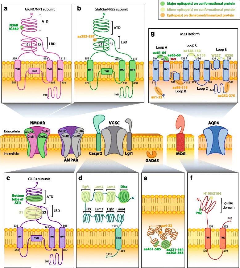
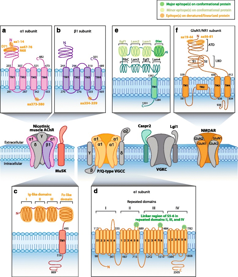
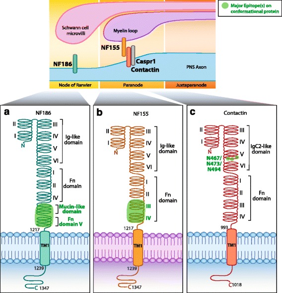
Main body: The purpose of this review is to discuss the mapping of the epitope targets of autoantibodies in CNS and PNS antibody-mediated disorders, such as N-methyl-D-aspartate receptor (NMDAR), α-amino-3-hydroxy-5-methyl-4-isoxazolepropionic acid receptor (AMPAR), leucine-rich glioma-inactivated protein 1 (Lgi1), contactin-associated protein-like 2 (Caspr2), myelin oligodendrocyte glycoprotein (MOG), aquaporin-4 (AQP4), 65 kDa glutamic acid decarboxylase (GAD65), acetylcholine receptor (AChR), muscle-specific kinase (MuSK), voltage-gated calcium channel (VGCC), neurofascin (NF), and contactin. We also address the methods used to analyze these epitopes, the relevance of their determination, and how this knowledge can inform studies on autoantibody pathogenicity. Furthermore, we discuss triggers of autoimmunity, such as molecular mimicry, ectopic antigen expression, epitope spreading, and potential mechanisms for the rising number of double autoantibody-positive patients.

Conclusions: Molecular insights into specificity and role of autoantibodies will likely improve diagnosis and treatment of CNS and PNS neuroimmune diseases.

Keywords: Autoantibody; CNS disorders; Epitope mapping; Epitope spreading; PNS disorders.


Figures

Fig. 1
Fig. 1
Epitopes of human CNS autoantibodies. Two subtypes of ionotropic glutamate receptors (iGluRs) are the N-methyl-D-aspartate receptor (NMDAR) and the α-amino-3-hydroxy-5-methyl-4-isoxazolepropionic acid receptor (AMPAR). Subunits of NMDAR (a, b) and AMPAR (c) contain an extracellular amino terminal domain (ATD) and a ligand-binding domain (LBD) formed by segment 1 (S1) and segment 2 (S2). Anti-NMDAR antibodies recognize amino acid (aa) N368 and G369 which reside on the bottom lobe in the ATD of the GluN1 subunit (a, Q05586) [35]. Anti-NMDAR antibodies also bind to an epitope at aa283-287 within S1 of the LBD in subunits GluN2a and GluN2b (b, Q12879; only GluN2A is shown) [67]. Anti-AMPAR antibodies are directed against an extracellular epitope within the bottom lobe in the ATD of the GluR1 subunit (c, P42261), but specific aa in the ATD have not been mapped [32]. d Contactin-associated protein 2 (Caspr2, Q9UHC6) consists of eight domains and forms part of the voltage-gated potassium channel (VGKC) complex with the leucine-rich glioma-inactivated protein 1 (Lgi1). Anti-Caspr2 antibodies recognize the extracellular N-terminal half (domains I–IV; discoidin (Disc), lamininG (Lam)-1, Lam2, and epidermal growth factor 1 (Egf1), respectively), but most commonly bind to an epitope within the Disc domain [37, 38]. e Anti-cytoplasmic enzyme glutamic acid decarboxylase 65 (GAD65) antibodies recognize an epitope at aa221-444 [48], aa451-585, and aa308-365 [47]. Anti-GAD65 antibodies have also been shown to bind to linearized protein at aa4-22 [47]. f Myelin oligodendrocyte glycoprotein (MOG, Q16653) is a myelin protein expressed on oligodendrocytes. Anti-MOG antibodies recognize a epitopes at P42 and at H103/S104 within the immunoglobulin (Ig)-like domain [56]. g Aquaporin-4 isoform M23 (AQP4-M23) is a water channel expressed on astrocytes. Anti-AQP4-M23 antibodies recognize epitopes within loop C (aa146-150) and loop E (aa227-228), but mostly in loop A (aa61-64) [62]. An additional study identified extracellular epitopes within loop A (aa66-69), loop C (N153), and loop E (H230) [65]. Reported intracellular epitopes include aa1-22, aa88-113, and aa252-275 [61]. D69 (red) is vital in maintaining the conformational structure of loop A [63]. Human AQP4-M23 sequence is derived from [210, 211]. Human protein topology, i.e., aa sequences and transmembrane domains (TM), is adapted from UniProt database and UniProt identifiers are shown between brackets. Diagrams do not depict protein crystal structure. Green highlights indicate major (dark green) and minor (light green) epitopes mapped by methods which retain the native in vivo conformational structure of proteins, such as cell-based assays. Orange highlights epitopes determined by methods that denature or linearized proteins, such as western blots and ELISAs (Table 1)
Fig. 2
Fig. 2
Epitopes of human PNS autoantibodies. Nicotinic muscle acetylcholine receptor (AChR) and muscle-specific kinase (MuSK, O15146) are postsynaptic muscle proteins. Anti-AChR antibodies target an extracellular epitope on the α1 subunit within amino acids (aa)1-14 [79] and aa67-76 [33]. Cytoplasmic epitopes have also been mapped in the α1 subunit at aa373-380 (a, P02708) and aa354–359 in the β1 subunit (b, P11230) [82]. c MuSK contains three immunoglobulin (Ig)-like domains and a Frizzled (Fz)-like domain. Anti-MuSK antibodies bind to the three N-linked Ig-like domains and the Fz-like domain [–85]. d The neuronal P/Q-type voltage-gated calcium channel (VGCC) consists of four repeated domains, each containing six segments (S), with a linker region between S5 and S6. Anti-P/Q-type VGCC antibodies recognize epitopes in the linker region of repeating domains II–IV [–96]. e Neuronal anti-contactin-associated protein 2 (Caspr2, Q9UHC6) antibodies recognize the extracellular N-terminal half (domains I–IV) but most commonly bind to an epitope within the discoidin (Disc) domain [37, 38]. f Neuronal N-methyl-D-aspartate receptor (NMDAR) contains an extracellular amino terminal domain (ATD) and a ligand-binding domain (LBD) formed by segment 1 (S1) and segment 2 (S2). Anti-NMDAR antibodies recognize aa19-44 and aa56-81 in the ATD of the GluN1 subunit (f, Q05586) [72]. Human protein topology, i.e., aa sequences and transmembrane domains (TM), is adapted from UniProt database, and UniProt identifiers are shown between brackets. Diagrams do not depict protein crystal structure. Green highlights major (dark green) and minor (light green) epitopes mapped by methods which retain the native in vivo conformational structure of the protein, such as cell-based assays. Orange highlights epitopes determined by methods which denature protein, such as western blots and ELISAs (Table 1)
Fig. 3
Fig. 3
Epitopes of human PNS autoantibodies at the node of Ranvier. There are two isoforms of neurofascin (NF). a The neuronal isoform NF 186 (NF186, O94856) that resides within the node. b The glial isoform NF 155 (NF155, O94856) that resides on the myelin loop within the paranode. Anti-NF186 antibodies target the extracellular fibronectin type III (Fn) V (or Fn5) and Mucin-like domain. Anti-NF155 antibodies target extracellular domains Fn III (or Fn3) and Fn IV (Fn4) [107]. c Antibodies directed against neuronal contactin (Q12860) target N-glycosylation sites N467, N473, and N494, within the extracellular immunoglobulin (Ig) C2-like domain 5 [108]. Human protein topology, i.e., aa sequences and transmembrane domains (TM), is adapted from UniProt database, and UniProt identifiers are shown between brackets. Diagrams do not depict protein crystal structure. Dark green highlights major epitopes mapped by methods which retain the native in vivo conformational structure of the protein, such as cell-based assays (Table 1)

References

    1. Irani SR, Gelfand JM, Al-Diwani A, Vincent A. Cell-surface central nervous system autoantibodies: clinical relevance and emerging paradigms. Ann Neurol. 2014;76:168–184. doi: 10.1002/ana.24200. - DOI - PMC - PubMed
    1. McKeon A, Pittock SJ. Paraneoplastic encephalomyelopathies: pathology and mechanisms. Acta Neuropathol. 2011;122:381–400. doi: 10.1007/s00401-011-0876-1. - DOI - PubMed
    1. Vincent A, Bien CG, Irani SR, Waters P. Autoantibodies associated with diseases of the CNS: new developments and future challenges. Lancet Neurol. 2011;10:759–772. doi: 10.1016/S1474-4422(11)70096-5. - DOI - PubMed
    1. Coutinho E, Harrison P, Vincent A. Do neuronal autoantibodies cause psychosis? A neuroimmunological perspective. Biol Psychiatry. 2014;75:269–275. doi: 10.1016/j.biopsych.2013.07.040. - DOI - PubMed
    1. Mohammad SS, Ramanathan S, Brilot F, Dale RC. Autoantibody-associated movement disorders. Neuropediatrics. 2013;44:336–345. doi: 10.1055/s-0033-1358603. - DOI - PubMed

Publication types

MeSH terms

LinkOut - more resources