. 2016 Oct 15;129(20):3859-3867.
doi: 10.1242/jcs.190322. Epub 2016 Aug 30.

Endo-lysosomal TRP mucolipin-1 channels trigger global ER Ca2+ release and Ca2+ influx

Affiliations

Endo-lysosomal TRP mucolipin-1 channels trigger global ER Ca2+ release and Ca2+ influx

Bethan S Kilpatrick et al. J Cell Sci. .

Abstract

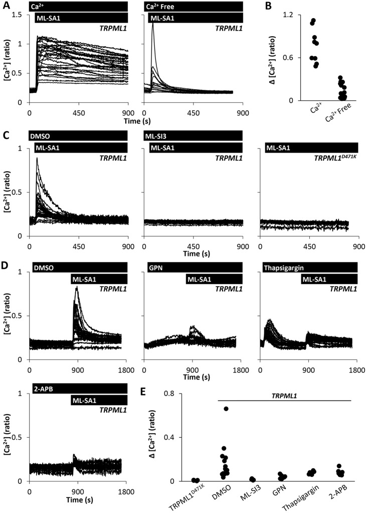
Transient receptor potential (TRP) mucolipins (TRPMLs), encoded by the MCOLN genes, are patho-physiologically relevant endo-lysosomal ion channels crucial for membrane trafficking. Several lines of evidence suggest that TRPMLs mediate localised Ca2+ release but their role in Ca2+ signalling is not clear. Here, we show that activation of endogenous and recombinant TRPMLs with synthetic agonists evoked global Ca2+ signals in human cells. These signals were blocked by a dominant-negative TRPML1 construct and a TRPML antagonist. We further show that, despite a predominant lysosomal localisation, TRPML1 supports both Ca2+ release and Ca2+ entry. Ca2+ release required lysosomal and ER Ca2+ stores suggesting that TRPMLs, like other endo-lysosomal Ca2+ channels, are capable of 'chatter' with ER Ca2+ channels. Our data identify new modalities for TRPML1 action.

Keywords: Ca2+; Endoplasmic reticulum; Lysosomes; TRP channels.


Conflict of interest statement

The authors declare no competing or financial interests.

Figures

Fig. 1.
Fig. 1.
The TRP mucolipin agonist ML-SA1 evokes global Ca2+ signals. (A–D) Effect of ML-SA1 on cytosolic Ca2+ levels. Cytosolic Ca2+ levels of individual Fura-2-loaded HeLa cells (A) or fibroblasts (B) from an exemplar population stimulated with increasing concentrations of ML-SA1. (C,D) Summary data quantifying the peak response (C) and percentage of responsive cells within the population (D). Each data point represents an independent repetition. A total of 350–788 HeLa cells and 81–140 fibroblasts were analysed. (E,F) Effect of the mucolipin inhibitor ML-SI3 on agonist-evoked Ca2+ signals. (E) Cytosolic Ca2+ levels of HeLa cells stimulated with 20 µM ML-SA1 in the presence of vehicle (0.1% DMSO) or 10 µM ML-SI3. (F) Summary data for the experiments in E (366–406 cells).
Fig. 2.
Fig. 2.
Subcellular distribution of TRPML1. (A) Confocal fluorescence images (green) of fixed HeLa cells expressing GFP-tagged LAMP1 (left), TRPML1 (middle) or TRPML1D471K (right). Nuclei were stained using DAPI (blue). (B,C) Confocal fluorescence images of cells expressing GFP–TRPML1 or GFP–TRPML1D471K and either colabelled with Lysotracker® Red (LTR) and imaged live (B) or co-transfected with LAMP1-mRFP and imaged following fixation (C). Overlays of images are shown in the right panels where arrows delineate the regions from which red–green intensity plots were derived. Scale bar: 10 µm. (D,E) Summary data of experiments in C quantifying Pearson's correlation coefficients (D) and the percentage of vesicles showing colocalisation (E). Each point represents an individual cell from two independent transfections.
Fig. 3.
Fig. 3.
Agonist-evoked Ca2+ signals require TRPML1. (A–C) Effect of TRPML expression on ML-SA1-evoked Ca2+ signals. (A) Cytosolic Ca2+ levels of individual LAMP1, TRPML1- or TRPML1D471K-expressing HeLa cells stimulated with 20 µM ML-SA1. (B,C) Summary data of experiments in A where each data point represents an independent repetition. The total number of cells analysed was 193–367. (D,E) Effect of TRPML expression on MK6-83-evoked Ca2+ signals. (D) Cytosolic Ca2+ levels of individual TRPML1- or TRPML1D471K-expressing HeLa cells stimulated with 20 µM MK6-83. (E) Summary data of experiments in D (111–128 cells). (F–I) Effect of ML-SI3 on agonist-evoked Ca2+ signals. Cytosolic Ca2+ levels of individual TRPML1-expressing HeLa cells stimulated with 20 µM ML-SA1 (F) or 20 µM MK6-83 (H) in the presence of vehicle (0.1% DMSO) or 10 µM ML-SI3. Summary data are shown in panels (G; 120 cells) and (I; 101–107 cells), respectively.
Fig. 4.
Fig. 4.
TRPML1 couples lysosomal and ER Ca2+ release. (A,B) Effect of extracellular Ca2+ on agonist-evoked Ca2+ responses. (A) Cytosolic Ca2+ levels of TRPML1-expressing HeLa cells stimulated with 20 µM ML-SA1 in Ca2+-containing or Ca2+-free Hepes-buffered saline. (B) Summary data of experiments in A where each data point represents an independent repetition. The total number of cells analysed was 184–284. (C) Cytosolic Ca2+ levels of TRPML1 or TRPML1D471K-expressing HeLa cells stimulated with 20 µM ML-SA1 with or without 10 µM ML-SI3 in Ca2+-free medium. (D,E) Effect of disrupting Ca2+ stores on agonist-evoked Ca2+ responses. (D) Cytosolic Ca2+ levels of TRPML1-expressing HeLa cells stimulated with 20 µM ML-SA1 following pre-treatment with DMSO, 200 µM GPN, 1 µM thapsigargin or 100 µM 2-APB. (E) Summary data of experiments in D (71–462 cells). Experiments were performed in Ca2+-free medium.
Fig. 5.
Fig. 5.
TRPML1 mediates Ca2+ influx. (A,B) Effect of blocking store-operated Ca2+ influx on agonist-evoked Ca2+ responses. (A) Cytosolic Ca2+ levels of TRPML1-expressing HeLa cells stimulated with 20 µM ML-SA1 in the presence of vehicle (0.1% DMSO) or 20 µM BTP2. (B) Summary data of experiments in A where each data point represents an independent repetition. The total number of cells analysed was 151–173. (C–J) Effect of TRPML1 activation on Fe ion entry. HeLa cells expressing TRPML1 were stimulated with 20 µM ML-SA1 (C) or MK6-83 (E) and then 1 mM FeCl2 in the presence of vehicle (0.1% DMSO) or 10 µM ML-SI3. HeLa cells expressing TRPML1D471K were stimulated with either 20 µM ML-SA1 or MK6-83 (G). HeLa cells expressing TRPML1 were stimulated with 20 µM ML-SA1 and either 1 mM FeCl2 or 1 mM FeCl3 (I). Experiments were performed in nominally Ca2+-free medium. Fluorescence signals (denoted F) were recorded after excitation at the isosbestic wavelength of Fura-2 (360 nm) and normalised to that at the beginning of recording (denoted F0). Summary data (D,F,H,J; for C,E,G and I, respectively) quantifying the fractional loss of fluorescence 144s after addition of Fe2+ or Fe3+ under the various conditions (80–169 cells).

References

    1. Bae M., Patel N., Xu H., Lee M., Tominaga-Yamanaka K., Nath A., Geiger J., Gorospe M., Mattson M. P. and Haughey N. J. (2014). Activation of TRPML1 clears intraneuronal Aβ in preclinical models of HIV infection. J. Neurosci. 34, 11485-11503. 10.1523/JNEUROSCI.0210-14.2014 - DOI - PMC - PubMed
    1. Bargal R., Avidan N., Ben-Asher E., Olender Z., Zeigler M., Frumkin A., Raas-Rothschild A., Glusman G., Lancet D. and Bach G. (2000). Identification of the gene causing mucolipidosis type IV. Nat. Genet. 26, 118-123. 10.1038/79095 - DOI - PubMed
    1. Berridge M. J., Bootman M. D. and Roderick H. L. (2003). Calcium signalling: dynamics, homeostasis and remodelling. Nat. Rev. Mol. Cell Biol. 4, 517-529. 10.1038/nrm1155 - DOI - PubMed
    1. Brailoiu E., Churamani D., Cai X., Schrlau M. G., Brailoiu G. C., Gao X., Hooper R., Boulware M. J., Dun N. J., Marchant J. S. et al. (2009). Essential requirement for two-pore channel 1 in NAADP-mediated calcium signaling. J. Cell Biol. 186, 201-209. 10.1083/jcb.200904073 - DOI - PMC - PubMed
    1. Calcraft P. J., Ruas M., Pan Z., Cheng X., Arredouani A., Hao X., Tang J., Rietdorf K., Teboul L., Chuang K.-T. et al. (2009). NAADP mobilizes calcium from acidic organelles through two-pore channels. Nature 459, 596-600. 10.1038/nature08030 - DOI - PMC - PubMed

Publication types

MeSH terms