. 2016 Oct 15;418(2):242-7.
doi: 10.1016/j.ydbio.2016.08.029. Epub 2016 Aug 27.

Knockdown of the pericellular matrix molecule perlecan lowers in situ cell and matrix stiffness in developing cartilage

Affiliations

Knockdown of the pericellular matrix molecule perlecan lowers in situ cell and matrix stiffness in developing cartilage

Xin Xu et al. Dev Biol. .

Abstract

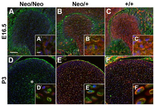
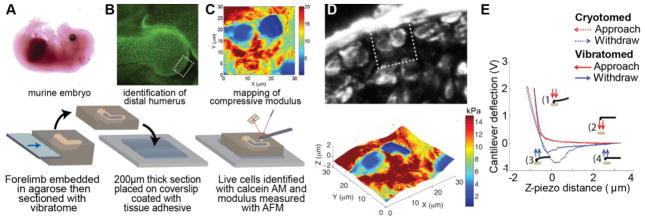
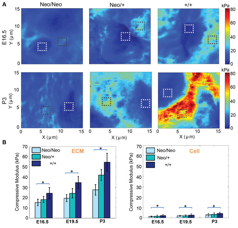
The pericellular matrix (PCM) is a component of the extracellular matrix that is found immediately surrounding individual chondrocytes in developing and adult cartilage, and is rich in the proteoglycan perlecan. Mutations in perlecan are the basis of several developmental disorders, which are thought to arise from disruptions in the mechanical stability of the PCM. We tested the hypothesis that defects in PCM organization will reduce the stiffness of chondrocytes in developing cartilage by combining a murine model of Schwartz-Jampel syndrome, in which perlecan is knocked down, with our novel atomic force microscopy technique that can measure the stiffness of living cells and surrounding matrix in embryonic and postnatal tissues in situ. Perlecan knockdown altered matrix organization and significantly decreased the stiffness of both chondrocytes and interstitial matrix as a function of age and genotype. Our results demonstrate that the knockdown of a spatially restricted matrix molecule can have a profound influence on cell and tissue stiffness, implicating a role for outside-in mechanical signals from the PCM in regulating the intracellular mechanisms required for the overall development of cartilage.

Keywords: Atomic force microscopy; Chondrocyte; Mechanobiology; Pericellular matrix.


Conflict of interest statement

The authors declare no competing interests.

Figures

Figure 1
Figure 1. The chondrocyte PCM in the developing humerus is disrupted by perlecan knockdown
Cryosections from mice homozygous (A, D), heterozygous (B, E) or wild-type (C, F) for the mutation in Hspg2 were stained for PCM components perlecan (red) and type VI collagen (green). Homozygous animals showed low levels of perlecan that appeared to be restricted to the intracellular compartment (A′, D′). In heterozygous and wild-type E16.5 embryos, perlecan was found throughout the developing cartilage, (B – C′). By P3, cell density decreased and perlecan became more restricted to the PCM in heterozygous and wild-type animals (E – F′). Type VI collagen reactivity was reduced in homozygous animals and was relatively disorganized in the center of the tissue (*, D). Blue = nuclei; bars: A = 75μm; A′ = 5μm.
Fig. 2
Fig. 2
AFM on vibratome sections enables the study of the biomechanics of viable cells and the surrounding ECM. (A) Forelimbs from freshly harvested murine embryos and pups were vibratomed to 200 μm thick. (B) All stiffness measurements were obtained proximal to the developing articular surface of the humerus (boxed region). Calcein-AM staining reveals cell viability is maintained throughout the forelimb. (C) Compressive modulus was measured the day of harvest via AFM. (D) Comparison of type VI collagen distribution and a surface height-stiffness map of the section shown in C (boxed region) indicates that stiffness of vibratomed samples is not dependent upon local topography. E) Representative force - displacement curves demonstrate that the stiffness of developing cartilage is dependent on whether the sections are vibratomed or cryotomed, see also (Xu et al., 2016).
Figure 3
Figure 3. Perlecan knockdown significantly decreases the stiffness of the cells and ECM in developing articular cartilage
A) High-resolution stiffness maps show an increase in stiffness with age and perlecan incorporation. Boxes indicate representative regions where cell (white) and ECM (black) stiffness were measured. B) Both cell and ECM stiffness are influenced by perlecan content (*p<0.0001). Two-way ANOVA analysis of ECM and cell stiffness reveals a significant effect of age (p<0.0001) and genotype (p<0.0001). The interaction between age and genotype was significant for both components of the tissue (p<0.01). Data for each time point were pooled from three independent litters in which all three genotypes were represented. Error bars = s.d.

References

    1. Alexopoulos LG, Youn I, Bonaldo P, Guilak F. Developmental and osteoarthritic changes in Col6a1-knockout mice: biomechanics of type VI collagen in the cartilage pericellular matrix. Arthritis Rheum. 2009;60:771–779. - PMC - PubMed
    1. Arikawa-Hirasawa E, Wilcox WR, Le AH, Silverman N, Govindraj P, Hassell JR, Yamada Y. Dyssegmental dysplasia, Silverman-Handmaker type, is caused by functional null mutations of the perlecan gene. Nat Genet. 2001;27:431–434. - PubMed
    1. Bischof JC, He X. Thermal stability of proteins. Ann NY Acad Sci. 2005;1066:12–33. - PubMed
    1. Buschmann MD, Kim YJ, Wong M, Frank E, Hunziker EB, Grodzinsky AJ. Stimulation of aggrecan synthesis in cartilage explants by cyclic loading is localized to regions of high interstitial fluid flow. Arch Biochem Biophys. 1999;366:1–7. - PubMed
    1. Calve S, Odelberg SJ, Simon HG. A transitional extracellular matrix instructs cell behavior during muscle regeneration. Dev Biol. 2010;344:259–271. - PMC - PubMed

Publication types

MeSH terms

LinkOut - more resources