. 2016 Sep 13;113(37):E5434-43.
doi: 10.1073/pnas.1521069113. Epub 2016 Aug 30.

Cd47-Sirpα interaction and IL-10 constrain inflammation-induced macrophage phagocytosis of healthy self-cells

Affiliations

Cd47-Sirpα interaction and IL-10 constrain inflammation-induced macrophage phagocytosis of healthy self-cells

Zhen Bian et al. Proc Natl Acad Sci U S A. .

Abstract

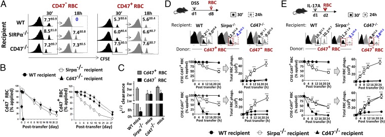
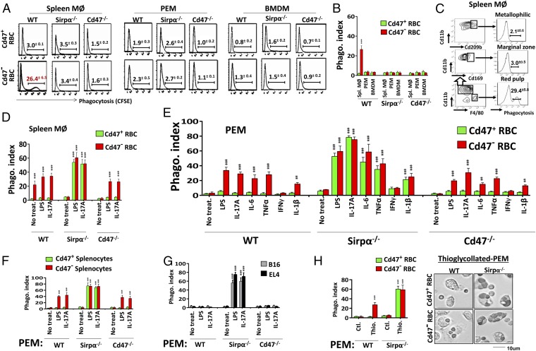
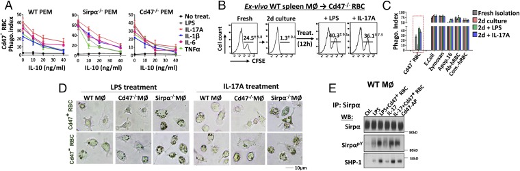
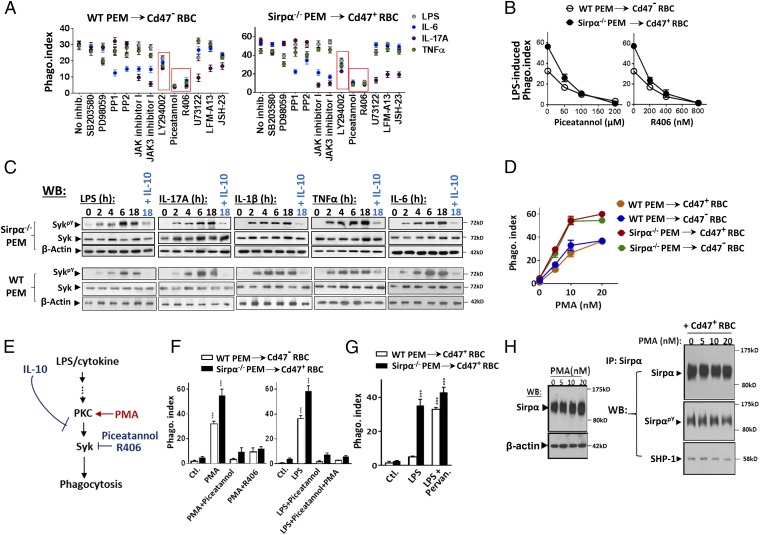
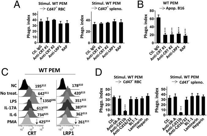
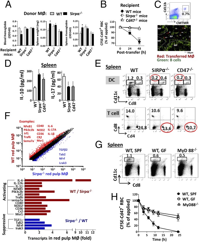
Rapid clearance of adoptively transferred Cd47-null (Cd47(-/-)) cells in congeneic WT mice suggests a critical self-recognition mechanism, in which CD47 is the ubiquitous marker of self, and its interaction with macrophage signal regulatory protein α (SIRPα) triggers inhibitory signaling through SIRPα cytoplasmic immunoreceptor tyrosine-based inhibition motifs and tyrosine phosphatase SHP-1/2. However, instead of displaying self-destruction phenotypes, Cd47(-/-) mice manifest no, or only mild, macrophage phagocytosis toward self-cells except under the nonobese diabetic background. Studying our recently established Sirpα-KO (Sirpα(-/-)) mice, as well as Cd47(-/-) mice, we reveal additional activation and inhibitory mechanisms besides the CD47-SIRPα axis dominantly controlling macrophage behavior. Sirpα(-/-) mice and Cd47(-/-) mice, although being normally healthy, develop severe anemia and splenomegaly under chronic colitis, peritonitis, cytokine treatments, and CFA-/LPS-induced inflammation, owing to splenic macrophages phagocytizing self-red blood cells. Ex vivo phagocytosis assays confirmed general inactivity of macrophages from Sirpα(-/-) or Cd47(-/-) mice toward healthy self-cells, whereas they aggressively attack toward bacteria, zymosan, apoptotic, and immune complex-bound cells; however, treating these macrophages with IL-17, LPS, IL-6, IL-1β, and TNFα, but not IFNγ, dramatically initiates potent phagocytosis toward self-cells, for which only the Cd47-Sirpα interaction restrains. Even for macrophages from WT mice, phagocytosis toward Cd47(-/-) cells does not occur without phagocytic activation. Mechanistic studies suggest a PKC-Syk-mediated signaling pathway, to which IL-10 conversely inhibits, is required for activating macrophage self-targeting, followed by phagocytosis independent of calreticulin Moreover, we identified spleen red pulp to be one specific tissue that provides stimuli constantly activating macrophage phagocytosis albeit lacking in Cd47(-/-) or Sirpα(-/-) mice.

Keywords: CD47; SIRPα; cytokine; macrophage; phagocytosis.


Conflict of interest statement

The authors declare no conflict of interest.

Figures

Fig. 1.
Fig. 1.
Inflammatory conditions induce acute anemia in mice deficient of the Cd47-Sirpα–mediated inhibition. (A) Generation of Sirpα KO mice. PCR genotyping shows that WT allele produces a DNA fragment of 228 bp, whereas mutated allele produces a fragment of 502 bp. Western blot (WB) confirmed depletion of Sirpα (∼120 kDa) protein in bone marrow leukocytes from Sirpα−/− mice. (B) Inducing colitis in mice by 1% or 2% DSS. Note that the colitic progression in Sirpα−/− mice induced by 1% DSS was comparable to that in WT induced with 2% DSS. (C and D) Acute anemia and splenomegaly developed in Sirpα−/− mice with colitis. WT mice and Sirpα−/− mice treated with DSS (2% DSS for WT and 1% DSS for Sirpα−/−) for 10 d were analyzed for peritoneal cavities (C) and peripheral blood (D). (E) H&E staining of spleens from WT and Sirpα−/− mice treated with 2% and 1% DSS for 10 d. (F) Time-course anemia development in Sirpα−/− mice during DSS-induced colitis and the correlation with IL-17 in serum. (G) IL-17 neutralization ameliorates anemia and splenomegaly in Sirpα−/− mice under colitis. An anti–IL-17 antibody (10 µg, i.v.) was given on days 6 and 8 (arrows) during DSS-induced colitis. (H) IL-17–containing colitic serum induces acute anemia in Sirpα−/− mice. Serum samples collected from healthy (ctl.) and colitic WT mice (2% DSS, 10 d) were administrated to healthy Sirpα−/− mice (i.v., 3×, arrows) with or without anti–IL-17 Ab. (I and J) IL-17A directly induces acute anemia and splenomegaly in Sirpα−/− mice and Cd47−/− mice. Healthy mice were given recombinant IL-17A (10 µg/kg, i.v.) on days 1 and 3 (arrows, J); anemia and splenomegaly were analyzed on day 5 (I) or in a time course manner (J). (K) Acute anemia and splenomegaly in Sirpα−/− and Cd47−/− mice under zymosan-peritonitis (3×, every other day), IL-6 administration (2×, 10 µg/kg, i.v.), LPS (0.25 mg/kg, i.p.), and CFA (1×, s.c.) administrations. Error bars are ±SEM **P < 0.01, ***P < 0.001 vs. control or the beginning time point. Data presented in each panel represent at least three independent experiments with n ≥ 4, if applicable.
Fig. 2.
Fig. 2.
Macrophage phagocytosis in vivo assessed by adoptive transfer experiments. (A–C) Clearance of adoptively transferred Cd47+ or Cd47 RBC in recipient mice. (A) FACS analyses of CFSE-RBCs in peripheral blood at 30 min (initial time point) and 18 h after transfer. (B) Time course clearance of CFSE-labeled RBCs. (C) The half-time (t1/2) of RBC clearance. Error bars are ±SEM. **P < 0.01, ***P < 0.001 vs. WT mice clearance of Cd47+ RBCs. (D) RBC clearance in mice under DSS-induced colitis. Mice treated with DSS (1% for Sirpα−/− mice, 2% for WT and Cd47−/− mice) for 8 d (d8) were transferred with CFSE-RBCs followed by determination of RBC clearance after 24 h. FACS data of Cd47+ RBCs and Cd47 RBCs in different mice at 30 min and 24 h after transfer were selectively shown. Total RBC phagocytosis was calculated based on the rates of CFSE-RBC clearance and the fact that Sirpα−/− mice eliminate both Cd47 RBC and Cd47+ RBC, whereas WT mice and Cd47−/− mice eliminate only Cd47 RBCs. (E) RBC clearance in mice treated with IL-17A. Mice treated once with IL-17A (10 µg/kg, i.v.) were transferred with CFSE-labeled Cd47+ or Cd47 RBCs a day later. Error bars are ±SEM. **P < 0.01, ***P < 0.001 vs. control or the initial time point. Data presented in each panel represent at least three independent experiments with n ≥ 4, if applicable.
Fig. 3.
Fig. 3.
Assaying macrophage phagocytosis ex vivo. (A and B) Macrophage phagocytosis toward RBCs. Freshly isolated splenic macrophages (MØ) and PEM and in vitro-derived BMDM were tested for phagocytosis toward CFSE-labeled Cd47+ or Cd47 RBCs. (C) Only red pulp macrophages are RBC phagocytes. (D) Activation of splenic MØ from Sirpα−/− and Cd47−/− mice for phagocytosis toward RBCs by LPS and IL-17A. (E) Activation of PEM for phagocytosis toward RBC by LPS, IL-6, IL-1β, IL-17, and TNFα, but not IFN-γ. (F and G) LPS and IL-17A–activated PEM phagocytosis toward splenocytes (F), B16, and EL4 (G). Error bars are ±SEM. **P < 0.01, ***P < 0.001 vs. no treatment controls. (H) Thioglycollate activates PEM phagocytosis toward RBCs. PEM lavaged without (ctl.) or with Brewer thioglycollate (3%, i.p.) elicitation was tested. Error bars are ±SEM. ***P < 0.001 vs. control PEM. Data presented in each panel represent at least three independent experiments with n ≥ 4, if applicable.
Fig. 4.
Fig. 4.
(A) IL-10 inhibits macrophage phagocytosis toward self. PEMs were treated with LPS and activating cytokines along with various concentrations of IL-10 before testing phagocytosis toward RBCs. (B) Phagocytic plasticity of WT red pulp macrophages. The phagocytic capacity toward Cd47 RBCs displayed by freshly isolated WT splenic macrophages was lost after 2 d (d2) of in vitro culturing. Treatments of cultured macrophages with LPS and IL-17A re-elicited their phagocytosis toward RBCs. (C) Phagocytosis toward Cd47 RBCs, E. coli, zymosan, apoptotic cells, and antibody or complement-bound hRBCs. Error bars are ±SEM. ***P < 0.001 vs. freshly isolated splenic macrophages. (D) Microscopic images of RBC phagocytosis by LPS- and IL-17A–treated splenic macrophages. (E) Sirpα ITIM phosphorylation and SHP-1 association under LPS and IL-17A treatments with or without Cd47 ligation. WT PEMs treated with LPS or IL-17A were further treated with Cd47+ RBCs or Cd47-AP (10 min, 37 °C) followed by cell lysis, Sirpα immunoprecipitation, and WB detection of Sirpα phosphorylation (SirpαpY) and SHP-1 association. Data presented in each panel represent at least three independent experiments with n ≥ 4, if applicable.
Fig. 5.
Fig. 5.
Role of Syk in phagocytic activation. (A) Testing cell signaling inhibitors on LPS/cytokine-induced macrophage phagocytic activation. PEMs were treated with LPS and cytokines in the presence of various inhibitors. After washing, inhibitor-free macrophages were tested for phagocytosis toward RBCs. (B) Syk inhibitors Piceatannol and R406 dose-dependently inhibited LPS-induced PEM phagocytic activation. (C) Syk activity is regulated by phagocytic stimuli and IL-10. PEMs treated with LPS and activating cytokines, or together with IL-10, were tested for total Syk and phosphorylated Syk (SykpY, specific at Y519/520). (D) PMA-induced macrophages phagocytosis toward RBCs. WT and Sirpα−/− PEMs were treated with PMA (37 °C, 30 min) before testing phagocytosis toward RBCs. (E) Depiction of PKC-Syk–mediated macrophage phagocytic activation toward self. (F) Syk is downstream of PKC. (Left) Inhibition of Syk by Piceatannol and R406 prevented PMA-induced phagocytic activation. (Right) PMA treatment failed to rescue LPS-mediated phagocytic activation-suppressed by Syk inhibition. Error bars are ±SEM. ***P < 0.001 vs. the respective controls. (G) Inhibition of SHP by pervanadate eliminates Cd47-dependent phagocytic recognition. LPS-treated WT PEMs were further briefly treated with pervanadate before testing for phagocytosis toward RBCs. Error bars are ±SEM. ***P < 0.001 vs. the respective controls. (H) PMA treatment does not affect Sirpα expression or Cd47 ligation-induced Sirpα phosphorylation and SHP-1 association. Data presented in each panel represent at least three independent experiments with n ≥ 4, if applicable.
Fig. 6.
Fig. 6.
(A) Blocking CRT or LRP1 failed to inhibit LPS/cytokine-induced PEM phagocytosis toward RBCs. LPS/cytokine-activated PEM phagocytosis toward RBCs was tested in the presence of anti-CRT antibody 1 (Abcam) and 2 (CST) (10 μg/mL each), anti-LRP1 (20 μg/mL), and the LRP1 receptor-associated protein (RAP; 20 μg/mL), all dialyzed free of sodium azide (note: sodium azide potently inhibits phagocytosis toward self-cells even at low concentrations). (B) CRT-LRP1 controls macrophage phagocytosis toward apoptotic cells. PEM (unstimulated) phagocytosis toward apoptotic B16 cells was tested in the presence of same antibodies and RAP as in A. Error bars are ±SEM. ***P < 0.001 vs. phagocytosis in the presence of control IgG. (C) Macrophage cell surface CRT or LRP1. PEMs, with or without (ctl.) treatment with LPS, IL-17A, IL-6, or PMA, were labeled for cell surface LRP1 and CRT followed by FACS. Increased and decreased expressions were marked by arrows. (D) Exploring other phagocytic receptors. Activated PEM phagocytosis toward RBCs was tested in the presence of antibodies against SR-A, Fc receptor Cd16/32 (10 μg/mL of each), inhibitors against SR-B (BLT1, 5 μM), dectin (laminarin, 100 μg/mL), complement (heparin, 40 U/mL), and antibody against Cd11b (10 μg/mL). Error bars are ±SEM. ***P < 0.001 vs. the respective control without inhibition. More data can be seen in SI Appendix, Figs. S10 and S11. Data presented in each panel represent at least three independent experiments with n ≥ 4, if applicable.
Fig. 7.
Fig. 7.
Sirpα−/− or Cd47−/− mice, but not WT mice, are deficient of macrophage stimulation in the spleen. (A–C) Monocytes/macrophages (2 × 10⁷) derived from bone marrow cells of WT or Sirpα−/− mice were labeled with CMTMR and transferred into three strains of recipient mice. (A) Only Sirpα−/− macrophages in WT mice displayed phagocytosis toward RBCs, resulting in anemia and splenomegaly. Error bars are ±SEM. *P < 0.05, **P < 0.01 vs. control by transferring WT monocytes/macrophages into WT mice. (B) Cotransfer of CFSE-labeled Cd47+ RBCs along with Sirpα−/− monocyte/macrophages into WT mice confirmed rapid RBC clearance. Error bars are ±SEM. ***P < 0.001 vs. the initial time point. (C) Distribution of CMTMR-labeled Sirpα−/− macrophages in spleen red pulp (RP) but not white pulp (WP). (D) Higher levels of IL-10 and lesser IL-17 produced by spleen cells and in serum from Sirpα−/− and Cd47−/− mice. (E) Decreases in Cd11c+Cd8 DC and Cd4+ helper (Th) lymphocytes in the spleen of Sirpα−/− and Cd47−/− mice. (F) Transcription profiling of red pulp macrophages from WT and Sirpα−/− mice. F4/80+ red pulp macrophages were affinity isolated before mRNA isolation and profiling. The red-colored molecules (most being activating) are expressed at higher levels in WT than in Sirpα−/− red pulp macrophages, whereas the blue-colored molecules (most being suppressive) are expressed oppositely. (G) Reduction of Cd11c+Cd8 DCs in the spleens of MyD88−/− mice and germ-free (GF)–conditioned mice. (H) Attenuated clearance of Cd47 RBC in MyD88−/− mice and GF mice. Data presented in each panel represent at least three independent experiments with n ≥ 4, if applicable.

References

    1. Oldenborg PA, et al. Role of CD47 as a marker of self on red blood cells. Science. 2000;288(5473):2051–2054. - PubMed
    1. Matozaki T, Murata Y, Okazawa H, Ohnishi H. Functions and molecular mechanisms of the CD47-SIRPalpha signalling pathway. Trends Cell Biol. 2009;19(2):72–80. - PubMed
    1. Barclay AN, Van den Berg TK. The interaction between signal regulatory protein alpha (SIRPα) and CD47: Structure, function, and therapeutic target. Annu Rev Immunol. 2014;32:25–50. - PubMed
    1. Chao MP, Weissman IL, Majeti R. The CD47-SIRPα pathway in cancer immune evasion and potential therapeutic implications. Curr Opin Immunol. 2012;24(2):225–232. - PMC - PubMed
    1. Barclay AN. Signal regulatory protein alpha (SIRPalpha)/CD47 interaction and function. Curr Opin Immunol. 2009;21(1):47–52. - PMC - PubMed

Publication types