

Intracranial haemorrhage in pregnancy
- PMID: 27579059
- PMCID: PMC4989666
- DOI: 10.1258/om.2009.090030
Intracranial haemorrhage in pregnancy
Abstract
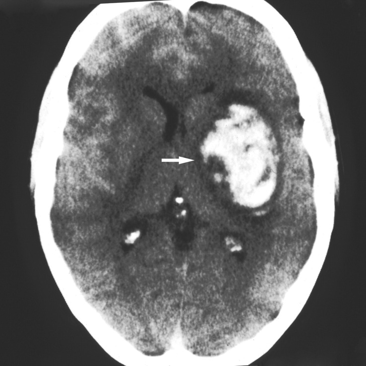
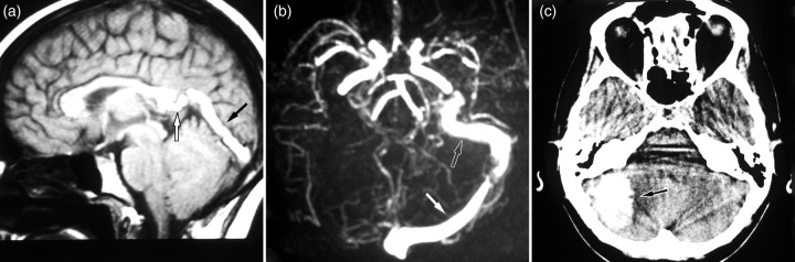
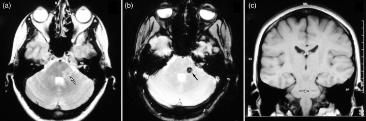
Intracranial haemorrhage (ICH) is a rare, yet potentially devastating event in pregnancy. There is a risk of maternal mortality or morbidity and a significant risk to the unborn child. The risk of haemorrhage increases during the third trimester and is greatest during parturition and the puerperium. ICH can be extradural, subdural, subarachnoid or intraparenchymal. Causes of bleeding include trauma, arteriovenous malformations, aneurysms, preeclampsia/eclampsia and venous thrombosis. Urgent neurosurgical conditions generally outweigh obstetric considerations in management decisions, although anaesthetic and surgical modifications can be made to minimize adverse effects to the fetus.
Keywords: arteriovenous malformations; cerebral aneurysms; intracerebral haemorrhage; pregnancy; stroke; subarachnoid haemorrhage.
Figures
References
-
- Jaigobin C, Silver FL. Stroke and pregnancy. Stroke 2000;31:2948–51 - PubMed
-
- Dias MS, Sekhar LN. Intracranial hemorrhage from aneurysms and arteriovenous malformations during pregnancy and the puerperium. Neurosurgery 1990;27:855–65; discussion 65–6 - PubMed
-
- Amias AG. Cerebral vascular disease in pregnancy. I. Haemorrhage. J Obstet Gynaecol Br Commonwealth 1970;77:100–20 - PubMed
-
- Wiebers DO, Whisnant JP. The incidence of stroke among pregnant women in Rochester, Minn, 1955 through 1979. JAMA 1985;254:3055–7 - PubMed
-
- Barno A, Freeman DW. Maternal deaths due to spontaneous subarachnoid hemorrhage. Am J Obstet Gynecol 1976;125:384–92 - PubMed
Publication types
LinkOut - more resources
Full Text Sources