

Laser Resection of Fibroepithelial Polyps with Digital Ureteroscopy
- PMID: 27579383
- PMCID: PMC4996577
- DOI: 10.1089/cren.2015.29014.sgh
Laser Resection of Fibroepithelial Polyps with Digital Ureteroscopy
Abstract
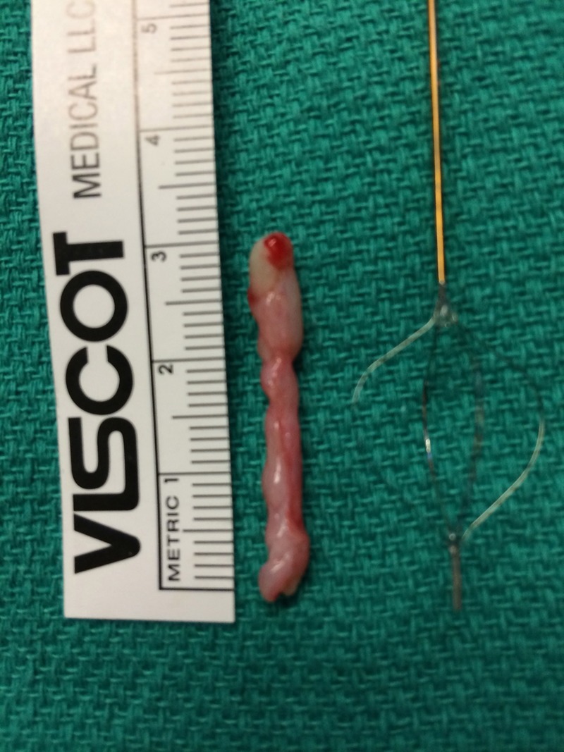
Fibroepithelial polyps (FEPs) are rare benign upper urinary-tract neoplasms originating from mesodermal components of the ureteral wall covered in normal urothelial epithelium. Historically, these lesions have been treated with endoscopic, laparoscopic, or open means depending on the size, number, and morphology of the polyps. Digital ureteroscopy (DURS) offers many advantages over fiber-optic endoscopy, including superior resolution and potential ergonomic benefits, given the absence of external cameras and light cords. We describe a case involving multiple proximal ureteral FEPs treated with flexible DURS and the holmium (Ho) laser, in which the digital ureteroscope offered exceptional visualization of the FEP stalks allowing for straightforward resection and endoscopic removal.
Figures


References
-
- Georgescu D, Multescu R, Geavlete BF, et al. . Fibroepithelial polyps—a rare pathology of the upper urinary tract. Rom J Morphol Embryol 2014;55:1325–1330 - PubMed
-
- Childs MA, Umbreit EC, Krambeck AE, et al. . Fibroepithelial polyps of the ureter: A single-institution experience. J Endourol 2009;23:1415–1419 - PubMed
-
- Zervas A, Rassidakis G, Nakopoulou L, et al. . Transitional cell carcinoma arising from a fibroepithelial ureteral polyp in a patient with duplicated urinary tract. J Urol 1997;157:2252–2253 - PubMed
-
- Andonian S, Okeke Z, Smith AD. Digital ureteroscopy: The next step. J Endourol 2008;22:603–605 - PubMed
Publication types
LinkOut - more resources
Full Text Sources
Other Literature Sources