

Two Unexpected Tumors in a Laparoscopic Nephrectomy Specimen, Including a Rare Tubulocystic Renal-Cell Carcinoma: A Case Report
- PMID: 27579398
- PMCID: PMC4996555
- DOI: 10.1089/cren.2015.0021
Two Unexpected Tumors in a Laparoscopic Nephrectomy Specimen, Including a Rare Tubulocystic Renal-Cell Carcinoma: A Case Report
Abstract
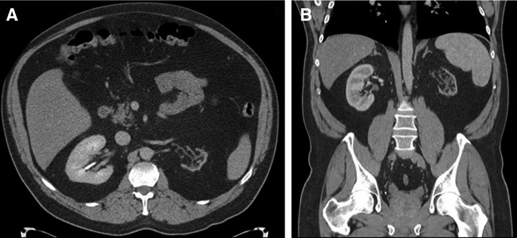
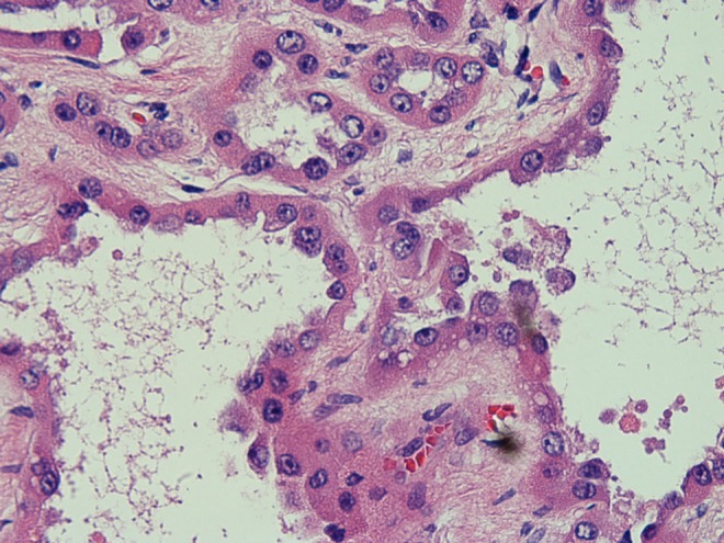
We present a case of a 52-year-old Caucasian male who underwent a laparoscopic nephrectomy for an atrophic kidney and was found to have two unexpected, synchronous kidney cancers. He had a remote history of testicular cancer complicated by lymphadenopathy and external ureteral compression. Over time, he developed an atrophic left kidney from obstructive uropathy. Years later, due to flank pain and renal scintigraphy showing minimal function, a laparoscopic nephrectomy was performed. Final pathology demonstrated papillary renal-cell carcinoma (RCC) and tubulocystic RCC. Tubulocystic RCC is a rare neoplasm thought to be an indolent subset of collecting duct carcinoma, but was identified as a unique entity in 2004. Currently, there are ∼100 cases of this neoplasm in the literature.
Figures

References
-
- Amin MB, Maclennan GT, Gupta R, et al. . Tubulocystic carcinoma of the kidney: Clinicopathologic analysis of 31 cases of a distinctive rare subtype of renal cell carcinoma. Am J Surg Pathol 2009;33:384–392 - PubMed
-
- Bhullar JS, Varshney N, Bhullar AK, Mittal VK. A new type of renal cancer-tubulocystic carcinoma of the kidney: A review of the literature. Int J Surg Pathol 2014;22:297–302 - PubMed
-
- Yang XJ, Zhou M, Hes O, et al. . Tubulocystic carcinoma of the kidney: Clinicopathologic and molecular characterization. Am J Surg Pathol 2008;32:177–187 - PubMed
-
- Mego M, Sycova-mila Z, Rejlekova K, et al. . Sunitinib in the treatment of tubulocystic carcinoma of the kidney. A case report. Ann Oncol 2008;19:1655–1656 - PubMed
-
- Maclennan GT, Farrow GM, Bostwick DG. Low-grade collecting duct carcinoma of the kidney: Report of 13 cases of low-grade mucinous tubulocystic renal carcinoma of possible collecting duct origin. Urology 1997;50:679–684 - PubMed
Publication types
LinkOut - more resources
Full Text Sources
Other Literature Sources