Review
. 2016 Dec 1;8(12):a028233.
doi: 10.1101/cshperspect.a028233.

Multiciliated Cells in Animals

Affiliations
Review

Multiciliated Cells in Animals

Alice Meunier et al. Cold Spring Harb Perspect Biol. .

Abstract

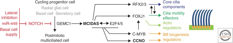
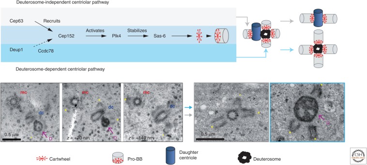
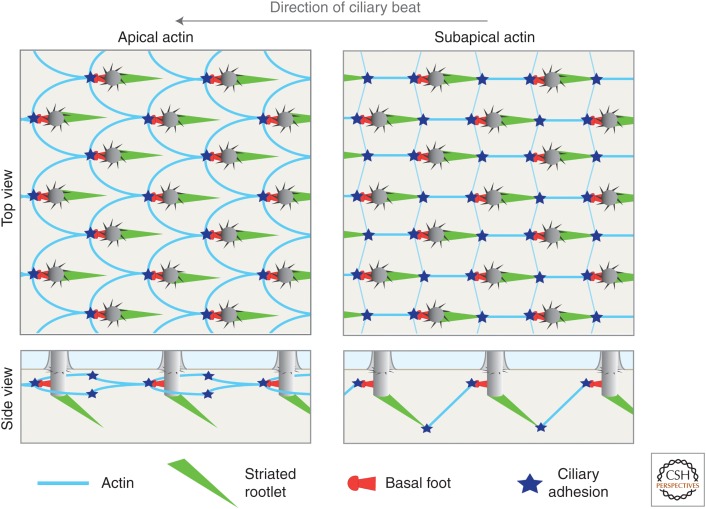
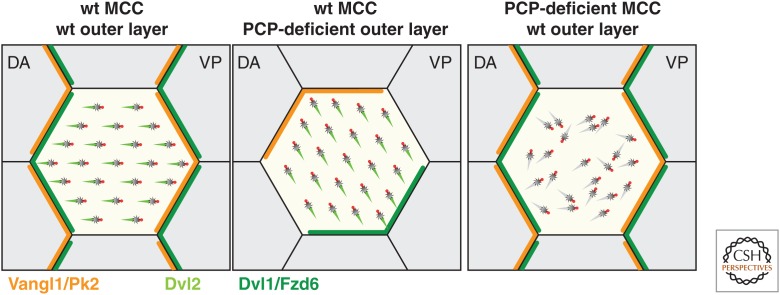
Many animal cells assemble single cilia involved in motile and/or sensory functions. In contrast, multiciliated cells (MCCs) assemble up to 300 motile cilia that beat in a coordinate fashion to generate a directional fluid flow. In the human airways, the brain, and the oviduct, MCCs allow mucus clearance, cerebrospinal fluid circulation, and egg transportation, respectively. Impairment of MCC function leads to chronic respiratory infections and increased risks of hydrocephalus and female infertility. MCC differentiation during development or repair involves the activation of a regulatory cascade triggered by the inhibition of Notch activity in MCC progenitors. The downstream events include the simultaneous assembly of a large number of basal bodies (BBs)-from which cilia are nucleated-in the cytoplasm of the differentiating MCCs, their migration and docking at the plasma membrane associated to an important remodeling of the actin cytoskeleton, and the assembly and polarization of motile cilia. The direction of ciliary beating is coordinated both within cells and at the tissue level by a combination of planar polarity cues affecting BB position and hydrodynamic forces that are both generated and sensed by the cilia. Herein, we review the mechanisms controlling the specification and differentiation of MCCs and BB assembly and organization at the apical surface, as well as ciliary assembly and coordination in MCCs.


Figures

Figure 1.
Figure 1.
Regulation of multiciliated cell (MCC) differentiation in vertebrates. MCC differentiation is triggered by Notch inhibition in radial glial cells of the ependyma, and in basal cells or secretory cells (trans-differentiation) in mucociliary epithelia. Notch inhibition triggers the activation of a regulatory cascade that involves the geminin-related proteins GEMC1 and MCIDAS, the transcription factors E2F4/5, RFX2/3, FOXJ1, and C-MYB, and the cyclin-like protein CCNO. This cascade triggers cell-cycle exit, basal body (BB) amplification, cytoskeleton remodeling, and ciliogenesis (see text for details).
Figure 2.
Figure 2.
Basal body (BB) amplification in mouse multiciliated cells (MCCs). (Upper panel) BB assembly occurs both through a deuterosome-dependent and a deuterosome-independent centriolar pathway. The deuterosome-independent pathway is initiated by the recruitment of CEP63 at centrosomal mother and daughter centrioles (for clarity, only the daughter centriole is represented). The deuterosome-dependent pathway is initiated by the recruitment of DEUP1 at the daughter centriole. Both pathways involve the same downstream molecular components for BB assembly, including CEP152, PLK4, and SAS-6 (Al Jord et al. 2014). Deuterosome-dependent assembly also involves the CCDC78 protein in Xenopus (Klos Dehring et al. 2013). (Left lower panel) Serial section electron micrographs showing pro-BB assembly in mouse ependymal MCCs. mc, mother centriole; dc, daughter centriole; D, deuterosome. Asterisks mark pro-BBs assembling near the centrosomal centrioles or around the deuterosome. (Right lower panel) Electron micrographs showing elongated pro-BBs (asterisks) around a centrosomal centriole (left) or a deuterosome (right).
Figure 3.
Figure 3.
A model illustrating the architecture of the actin cytoskeleton in Xenopus skin multiciliated cells (MCCs). The drawings represent the basal bodies (BBs) and either the actin apical or subapical pools in mature MCCs. This organization of the subapical network is proposed in Antoniades et al. (2014) and the model of apical actin organization is based on immunofluorescence data from the same study. Both actin pools are connected to the BBs by ciliary adhesions located in the vicinity of the basal feet (note that the exact localization—near or at the basal feet—remains to be determined) (Antoniades et al. 2014). In addition, actin bridges forming the subapical pool are connected to rootlet tips both along the direction of ciliary beat and perpendicular to this direction (Werner et al. 2011; Antoniades et al. 2014). For clarity, only the actin bridges connecting the BBs along the direction of ciliary beat are represented in the side view of the subapical actin network.
Figure 4.
Figure 4.
Multiciliated cell (MCC) polarization by the planar cell polarity (PCP) pathway in the Xenopus skin. Combined action of the PCP pathway and hydrodynamic forces align the BBs in a dorsoanterior (DA) to ventroposterior (VP) direction in wild-type (wt) Xenopus embryos. PCP components are asymmetrically distributed at the apical junctions, with a Dvl1/Fzd6 domain located upstream of flow and a Vangl1/Pk2 domain located downstream from flow (Butler and Wallingford 2015). When wt MCCs intercalate within a PCP-deficient outer epidermal layer in grafting experiments, intracellular basal body (BB) orientation is coordinated but improperly aligned with tissue polarity. When PCP-deficient MCCs intercalate within a wt outer layer, both intracellular and tissue-level polarity are lost (Mitchell et al. 2009). Note that in mouse ependymal and tracheal MCCs, the orientation of the Dvl/Fzd and Vangl/Pk domains with respect to the direction of ciliary beat is reversed (Vladar et al. 2012; Boutin et al. 2014).

References

    1. Adl SM, Simpson AG, Lane CE, Lukes J, Bass D, Bowser SS, Brown MW, Burki F, Dunthorn M, Hampl V, et al. 2012. The revised classification of eukaryotes. J Eukaryot Microbiol 59: 429–493. - PMC - PubMed
    1. Al Jord A, Lemaitre AI, Delgehyr N, Faucourt, Spassky N, Meunier A. 2014. Centriole amplification by mother and daughter centrioles differs in multiciliated cells. Nature 516: 104–107. - PubMed
    1. Almuedo-Castillo M, Salo E, Adell T. 2011. Dishevelled is essential for neural connectivity and planar cell polarity in planarians. Proc Natl Acad Sci 108: 2813–2818. - PMC - PubMed
    1. Amirav I, Wallmeier J, Loges NT, Menchen T, Pennekamp P, Mussaffi H, Abitbul R, Avital A, Bentur L, Dougherty GW, et al. 2016. Systematic analysis of CCNO variants in a defined population: Implications for clinical phenotype and differential diagnosis. Hum Mutat 37: 396–405. - PubMed
    1. Anderson RG. 1972. The three-dimensional structure of the basal body from the rhesus monkey oviduct. J Cell Biol 54: 246–265. - PMC - PubMed

LinkOut - more resources