Review
. 2016 Nov 1;8(11):a023630.
doi: 10.1101/cshperspect.a023630.

Potential Pathways of Abnormal Tau and α-Synuclein Dissemination in Sporadic Alzheimer's and Parkinson's Diseases

Affiliations
Review

Potential Pathways of Abnormal Tau and α-Synuclein Dissemination in Sporadic Alzheimer's and Parkinson's Diseases

Heiko Braak et al. Cold Spring Harb Perspect Biol. .

Abstract

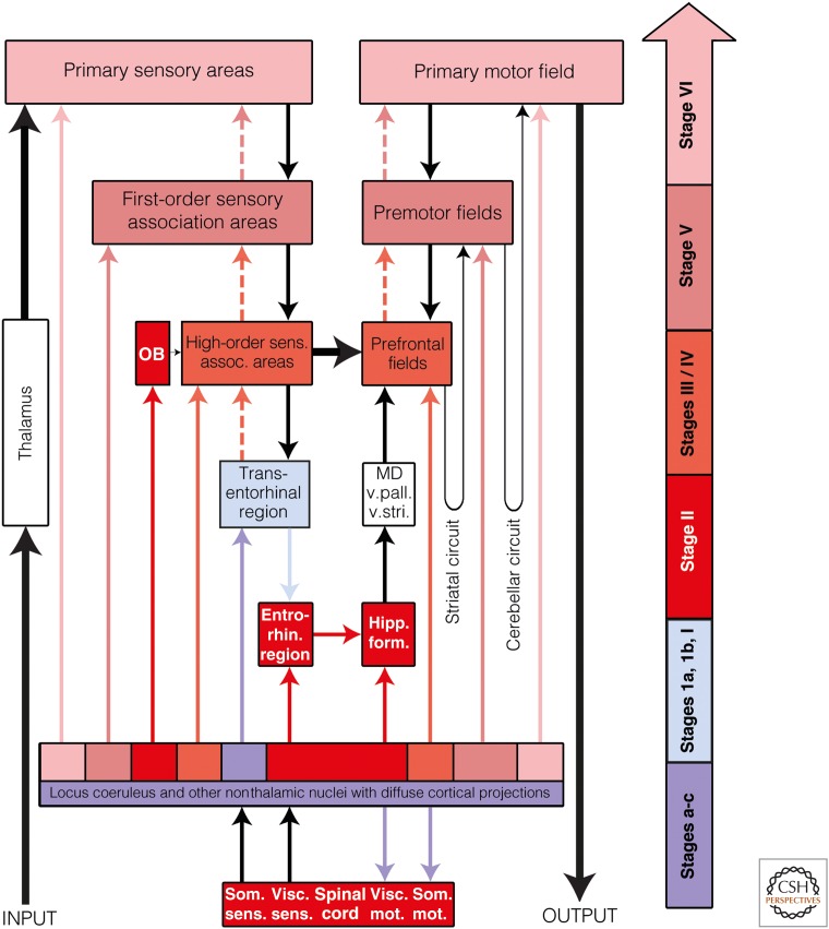
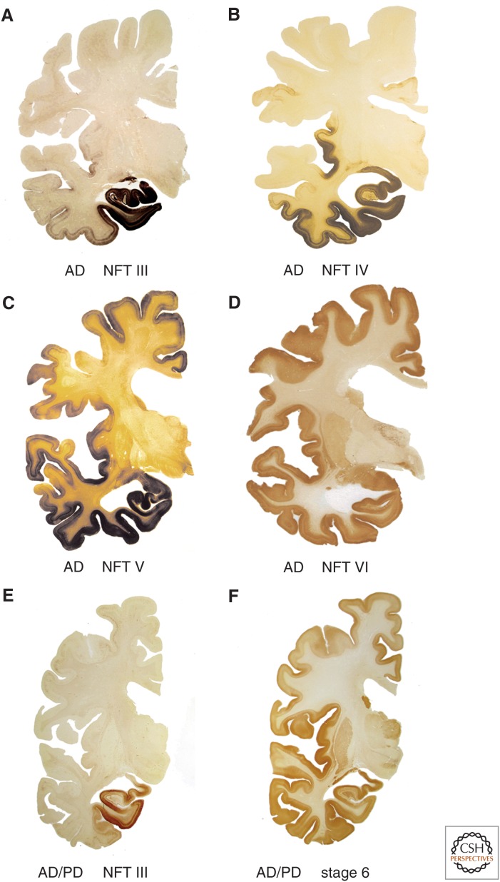
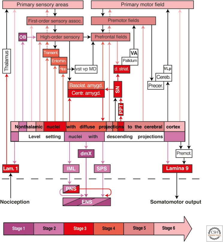
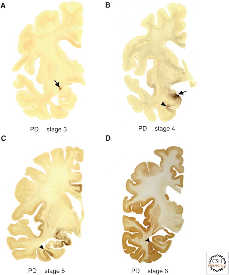
Experimental data indicate that transneuronal propagation of abnormal protein aggregates in neurodegenerative proteinopathies, such as sporadic Alzheimer's disease (AD) and Parkinson's disease (PD), is capable of a self-propagating process that leads to a progression of neurodegeneration and accumulation of prion-like particles. The mechanisms by which misfolded tau and α-synuclein possibly spread from one involved nerve cell to the next in the neuronal chain to induce abnormal aggregation are still unknown. Based on findings from studies of human autopsy cases, we review potential pathways and mechanisms related to axonal and transneuronal dissemination of tau (sporadic AD) and α-synuclein (sporadic PD) aggregates between anatomically interconnected regions.


Figures

Figure 1.
Figure 1.
Are small aggregates of pathological tau proteins transmissible in Alzheimer's disease (AD)? Summary of the stages of AD-related tau pathology (subcortical stages a–c, cortical pretangle stages 1a and 1b, and cortical neurofibrillary stages I–VI). (A) Axons of projection neurons of the locus coeruleus are the first structures to develop pretangle material immunoreactive against hyperphosphorylated tau protein (subcortical stage a). This aggregated material then fills the somatodendritic compartment (subcortical stage b). In subcortical stage c, additional nonthalamic nuclei with diffuse cortical projections (i.e., the upper raphe nuclei or the cholinergic magnocellular nuclei of the basal forebrain) begin to display pretangles. (B) The AD-related pathological process is confined to subcortical nuclei during stages a–c. (C) In pretangle stage 1a, pathological material develops in portions of nerve cell processes (possibly pathologically involved terminals of axons from the locus coeruleus) in the transentorhinal region of the cerebral cortex. This material subsequently fills pyramidal cells in the transentorhinal region (pretangle stage 1b). (DF) Drawings depict the systematic dissemination of the AD-related process from cortical pretangle stages 1a and 1b and neurofibrillary tangle (NFT) stages I and II (dark red) to neocortical high-order association areas at NFT stages III and IV (light red) and, finally, to first-order association areas and primary neocortical fields in NFT stages V and VI (pink). (From Braak and Del Tredici 2013; reprinted, with permission.)
Figure 2.
Figure 2.
Possible routes of tau transmission in Alzheimer's disease (AD). The color-coded arrow at the right indicates AD neuropathological stages a-VI and AD-related lesions. Color-coded arrows (excluding black arrows) are intended to indicate the routes by which the intraneuronal tau pathology may consecutively disseminate by anterograde transport. Directly above the block containing the locus coeruleus and other nonthalamic nuclei with diffuse projections to the cerebral cortex is a multicolored row: It is intended to signify the different groups of nerve cells belonging to these nuclei that become involved at various Parkinson's disease (PD) stages. OB, Olfactory bulb (chiefly the anterior olfactory nucleus); entorhin. region, entorhinal region; high-order sens. assoc. areas, high-order sensory association areas; hipp. form., hippocampal formation; MD, mediodorsal nuclei of the thalamus; som. mot., somatomotor; som. sens., somatosensory; visc. mot., visceromotor; visc. sens., viscerosensory; v. pall., ventral pallidum; v. stri., ventral striatum.
Figure 3.
Figure 3.
Neurofibrillary tangle (NFT) stages III–VI of sporadic Alzheimer's disease (AD) in 100 µm polyethylene glycol-embedded hemisphere sections. (A–D) During the limbic stages III and IV, areas of the anteromedial portion of the temporal lobe, including the transentorhinal region, entorhinal region, and hippocampal formation, become severely involved. From the transentorhinal region, the tau lesions enter (moving clockwise) into the adjoining high-order sensory association areas of the temporal neocortex (stage III) and from there into the medial temporal gyrus (stage IV). The insular cortex also becomes involved (stage IV). At neocortical stages V and VI, the premotor and first-order sensory association fields of the neocortex, and thereafter, the primary fields of the neocortex including the transverse gyrus of Heschl, develop very severe tau pathology. (E,F) Note that the transentorhinal region and the adjacent temporal allocortex are highly susceptible to both sporadic Parkinson's disease (PD) and AD. (A,B,D,E) AT8 immunohistochemistry, (C) Gallyas silver-iodide staining, and (F) α-synuclein immunohistochemistry.
Figure 4.
Figure 4.
Possible routes of α-synuclein transmission in Parkinson's disease (PD). The color-coded arrow at the bottom of the diagram indicates PD neuropathological stages 1–6 and PD-related lesions. Color-coded arrows (excluding black arrows) are intended to indicate the routes along which the pathology may disseminate transneuronally by anterograde or retrograde transport. A dashed black line separates the central from the peripheral and enteric nervous systems. The diagram shows the early involved level-setting nuclei with descending projections and nonthalamic nuclei with ascending projections in relationship to the pathological process. The lesions do not develop at all predilection sites within the nervous system simultaneously, but rather, sequentially. In stage 1 (dark purple), Lewy pathology is present within anterior olfactory structures (OB), in the dorsal motor nucleus of the vagus nerve (dmX), and in the intermediate reticular zone (not shown). The ambiguus nucleus is spared. Between stages 2 and 6, the pathological process progresses caudorostrally through the neuraxis. In stage 2 (light purple), the pathology reaches the level-setting nuclei of the lower brainstem (lower raphe group, gigantocellular nucleus of the reticular formation, and locus coeruleus), followed by a group of nonthalamic nuclei that project widely to the cerebral cortex (hypothalamic tuberomamillary nucleus, Meynert's basal nucleus, the interstitial nucleus of the diagonal band, medial septal nucleus). Involvement of midbrain nuclei (tegmental pedunculopontine nucleus [PPT], substantia nigra, pars compacta [SN], the upper raphe group) and the central subnucleus of the amygdala mark stage 3 (dark red). In stage 4 (dark red), additional subnuclei of the amygdala develop Lewy pathology and the cerebral cortex (transentorhinal region [transent], entorhinal region [entorhin], hippocampal formation [hipp]) become involved. During stages 5 and 6 (dark/light pink), lesions develop chiefly in projection neurons of the deep layers of the neocortex, beginning in the high-order sensory association areas and prefrontal fields. From there, the pathology advances into first-order sensory association areas and premotor fields, eventually reaching the primary sensory and motor fields. Spinal cord lesions, probably resulting from the level-setting nuclei via anterograde axonal transport, first develop at stage 2 in preganglionic sympathetic neurons of the intermediolateral nucleus (IML) and sacral parasympathetic nucleus (SPS) (light purple), followed during stage 3 by lesions in nociceptive projection neurons of layer 1 (lam. 1) and in α-motoneurons, including those of Onuf's nucleus, in layer 9 (lamina 9) (dark red). Alternatively, the IML could be reached by the spread of pathology from sympathetic postganglionic neurons of the peripheral celiac ganglion (peripheral nervous system [PNS]), which have contacts in the enteric nervous system (ENS). Based on known anatomical connectivities, the autonomic intramural plexus of the ENS could be either the source (dark purple, via retrograde transport) of the pathology in the dmX or the recipient (light purple, via anterograde transport) of the pathology from the dmX. Similarly, the lesions could disseminate to the locus coeruleus (stage 2) by retrograde transport from the olfactory bulb (OB) and/or from the dmX (stage 1). First-order sensory assoc., first-order sensory association areas; basolat. amygd., amygdala, basolateral subnuclei; OB, olfactory bulb (chiefly the anterior olfactory nucleus); high-order sensory, high-order sensory association areas; centr. amygd., central subnucleus, amygdala; cereb, cerebellum; dmX, dorsal motor nucleus of the vagal nerve; d. striat, dorsal striatum; ENS, enteric nervous system (submucosal and myenteric plexus); entorhin, entorhinal region; hipp, hippocampal formation (subiculum, Ammon's horn, and dentate fascia); MD, mediodorsal nuclei of the thalamus; PNS, peripheral nervous system (e.g., sympathetic trunk, celiac and superior cervical ganglia, and submandibular gland); precer, precerebellar nuclei; VA, ventral anterior nucleus of the thalamus; VLp, posterior ventrolateral nucleus of the thalamus; vp, ventral pallidum; vst, ventral striatum.
Figure 5.
Figure 5.
Lewy pathology in 100 µm polyethylene glycol–embedded hemisphere sections at stages 3–6 of sporadic Parkinson's disease (PD). (A) The arrow indicates the presence of the lesions in the central subnucleus of the amygdala. The cerebral cortex is still uninvolved at stage 3. (B) During stage 4, additional subnuclei of the amygdala develop Lewy pathology (the arrow points to cortical and basolateral subnuclei). Initial cortical lesions begin to appear at this stage in a specific portion of the anteromedial temporal lobe: the transentorhinal region (arrowhead). This region is a transitional zone wedged between allo- and neocortical regions and is especially developed only among higher primates, above all, in humans. (C) The pathological process progresses in stage 5 (moving clockwise from the transentorhinal region) into regions of the neocortex. Here, in addition to the increasing pathology present in the transentorhinal region (arrowhead), the inclusions in limbic portions of the temporal, insular, and cingulate cortex, in the hippocampal formation, and in the subnuclei of the amygdala gradually worsen. (D) During stage 6, the destruction in the temporal, insular, and cingulate regions is very severe. Not only the first-order sensory association areas and premotor fields of the neocortex, but also the neocortical primary sensory (including the primary auditory gyrus of Heschl) and motor fields become heavily involved. The arrowhead points to the transentorhinal region. (A–D) α-Synuclein immunohistochemistry.

References

    1. Adler CH, Dugger BN, Hinni ML, Lott DG, Driver-Dunckley E, Hidalgo J, Henry-Watson J, Serrano G, Sue LI, Nagel T, et al. 2014. Submandibular gland needle biopsy for the diagnosis of Parkinson disease. Neurology 82: 858–864. - PMC - PubMed
    1. Agnati LF, Bjekle B, Fuxe K. 1995. Volume versus wiring transmission in the brain: A new theoretical frame of neuropsychopharmacology. Med Res Rev 15: 33–45. - PubMed
    1. Aguzzi A. 2009. Beyond the prion principle. Nature 459: 924–925. - PubMed
    1. Aguzzi A, Calella AM. 2009. Prions: Protein aggregation and infectious diseases. Physiol Rev 89: 1105–1152. - PubMed
    1. Aguzzi A, Rajendran L. 2009. The transcellular spread of cytosolic amyloids, prions, and prionoids. Neuron 64: 783–790. - PubMed

MeSH terms