. 2016 Sep 1:6:32087.
doi: 10.1038/srep32087.

Calcineurin inhibitors cyclosporin A and tacrolimus protect against podocyte injury induced by puromycin aminonucleoside in rodent models

Affiliations

Calcineurin inhibitors cyclosporin A and tacrolimus protect against podocyte injury induced by puromycin aminonucleoside in rodent models

Xiujin Shen et al. Sci Rep. .

Abstract

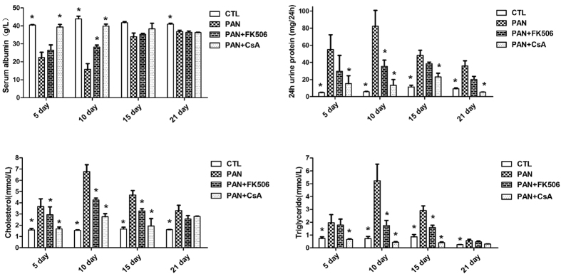
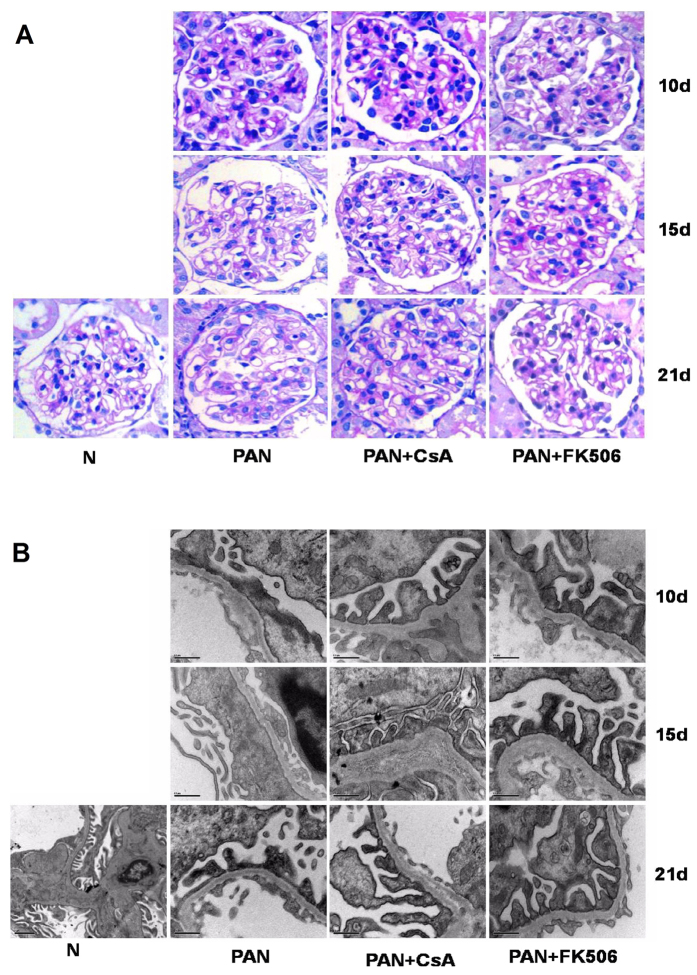
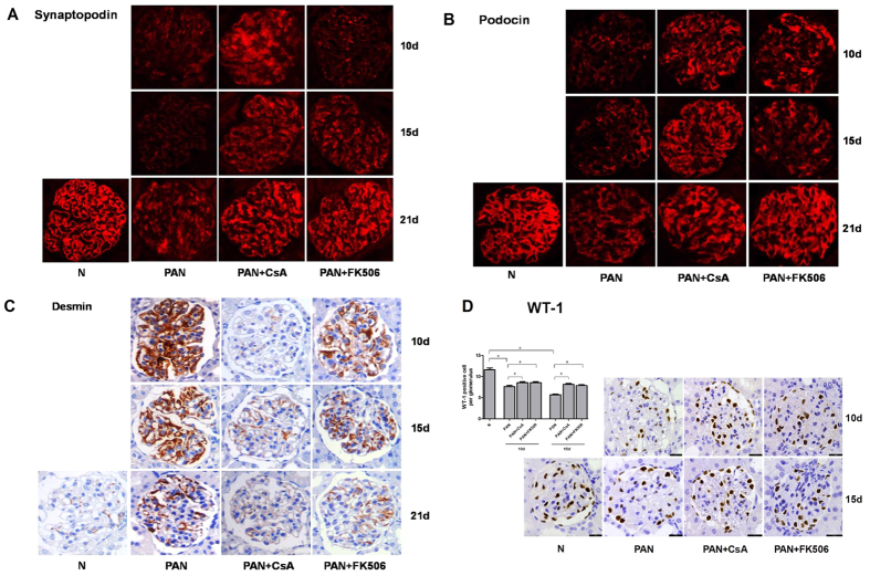
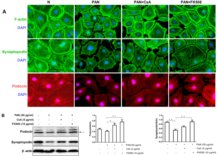
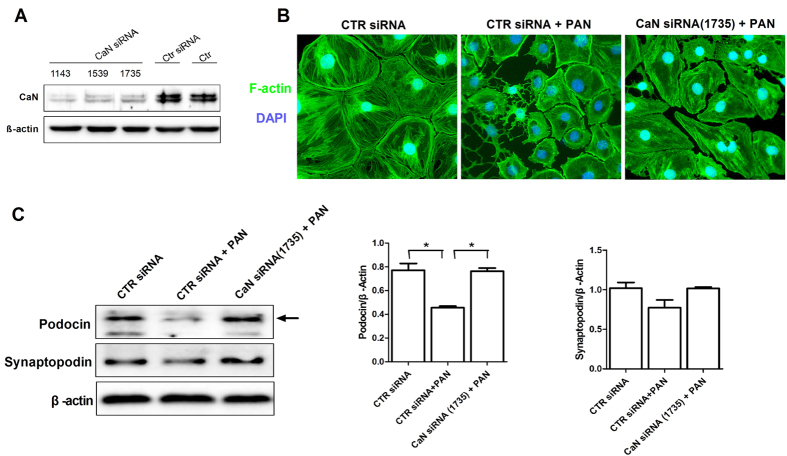
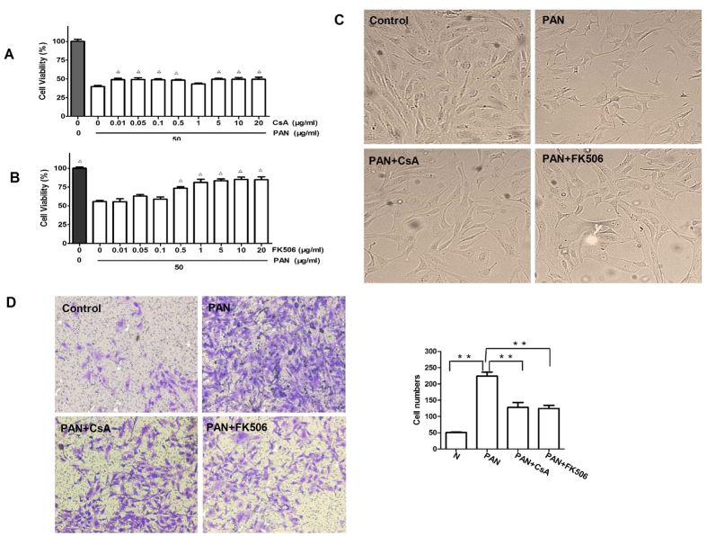
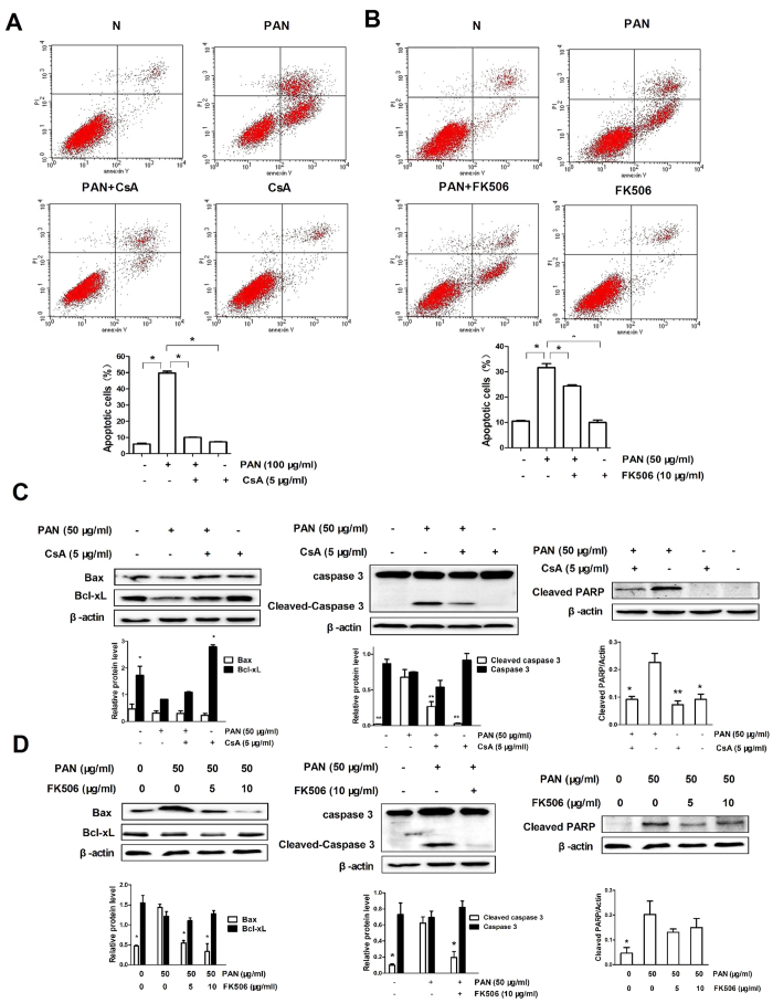
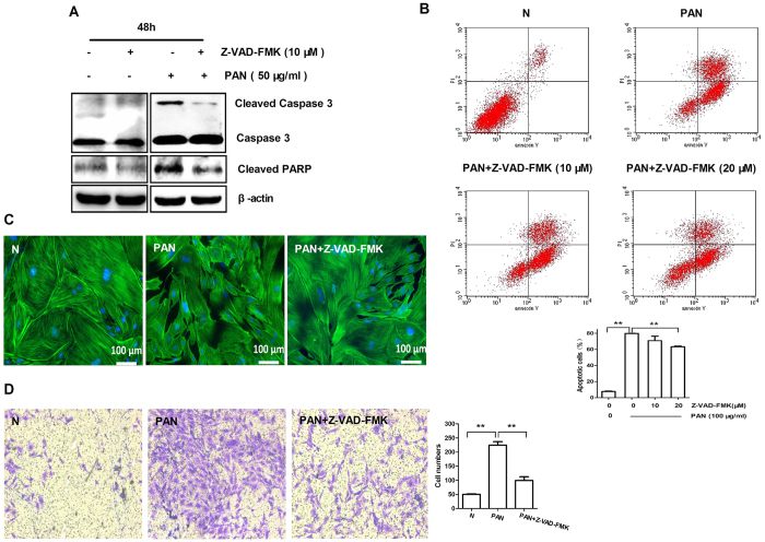
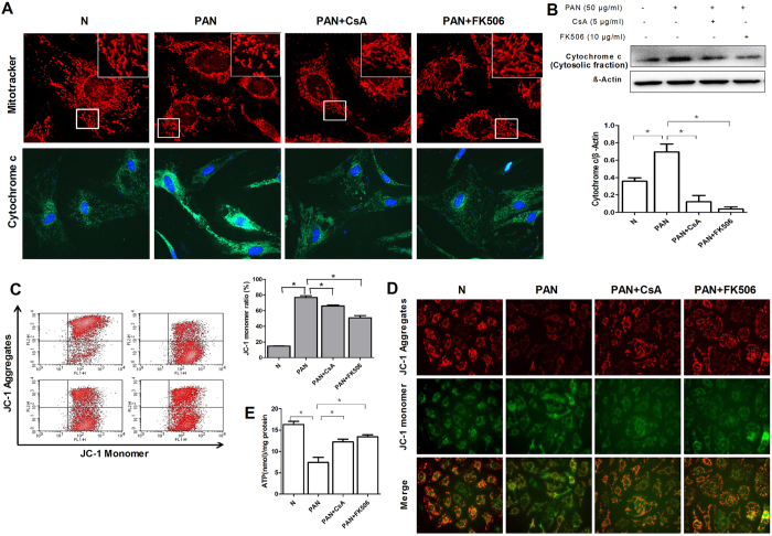
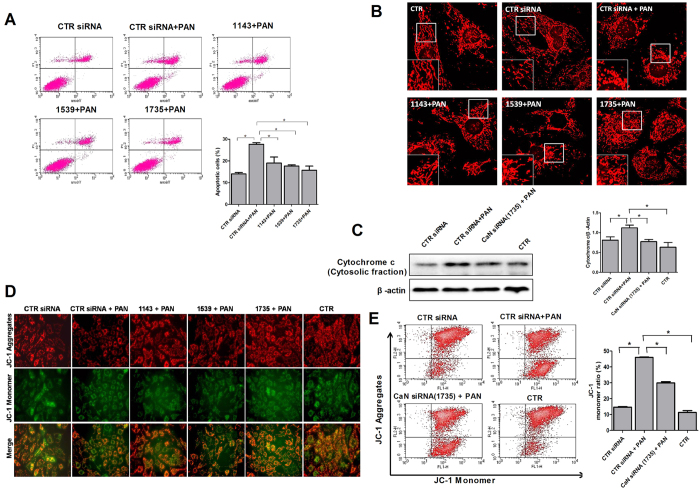
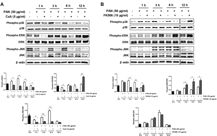
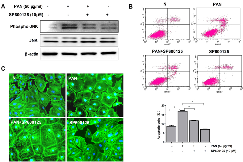
Podocyte injury and the appearance of proteinuria are features of minimal-change disease (MCD). Cyclosporin A (CsA) and tacrolimus (FK506) has been reported to reduce proteinuria in patients with nephrotic syndrome, but mechanisms remain unknown. We, therefore, investigated the protective mechanisms of CsA and FK506 on proteinuria in a rat model of MCD induced by puromycin aminonucleoside (PAN) and in vitro cultured mouse podocytes. Our results showed that CsA and FK506 treatment decreased proteinuria via a mechanism associated to a reduction in the foot-process fusion and desmin, and a recovery of synaptopodin and podocin. In PAN-treated mouse podocytes, pre-incubation with CsA and FK506 restored the distribution of the actin cytoskeleton, increased the expression of synaptopodin and podocin, improved podocyte viability, and reduced the migrating activities of podocytes. Treatment with CsA and FK506 also inhibited PAN-induced podocytes apoptosis, which was associated with the induction of Bcl-xL and inhibition of Bax, cleaved caspase 3, and cleaved PARP expression. Further studies revealed that CsA and FK506 inhibited PAN-induced p38 and JNK signaling, thereby protecting podocytes from PAN-induced injury. In conclusion, CsA and FK506 inhibit proteinuria by protecting against PAN-induced podocyte injury, which may be associated with inhibition of the MAPK signaling pathway.


Figures

Figure 1
Figure 1. CsA and FK506 ameliorate proteinuria, and serum albumin, triglyceride, and cholesterol abnormalities in SD rats.
CsA and FK506 reduced the 24-h urinary protein, decreased the triglyceride and cholesterol levels, and restored the serum albumin level in PAN-treated rats. CTL, normal rats; PAN, PAN-treated rats; PAN + FK506, intragastric administration of FK506 starting at the same time as PAN injection; PAN + CsA, intraperitoneal injection of CsA starting at the same time as PAN injection (n = 5 per group; *P < 0.05 vs PAN group).
Figure 2
Figure 2. Glomerular morphology and foot processes in normal, PAN, PAN + FK506, and PAN + CsA treated SD rats.
(A) PAS staining showed no difference in glomerular morphology between groups at indicated stages after CsA and FK506 treatment in PAN injured SD rats. Original magnification, X400. (B) Transmission electron microscopy showed extensive foot-process effacement at 10 and 15 days after PAN injection. CsA and FK506 treatment significantly decreased foot-process width compared with PAN-only rats. Scale bar, 0.5 μm (5 μm in normal rats). Images shown are representative from 5 rats for each treatment group.
Figure 3
Figure 3. Expression of synaptopodin, podocin, desmin and WT-1 in kidney glomerulus from SD rats subjected to various treatments.
Immunofluorescent and immunohistochemical staining for synaptopodin (A), podocin (B), desmin (C) and WT-1(D) showed that CsA treatments rescued the expression of synaptopodin, podocin and WT-1 in PAN-treated SD rats and inhibited PAN induced desmin expression. Original magnification, X400. Images shown are representative from 5 rats for each treatment group. WT-1 positive podocytes were calculated from more than 50 glomeruli (*P < 0.05).
Figure 4
Figure 4. CsA and FK506 pretreatment protect against PAN-induced injury in cultured mouse podocytes in vitro.
(A) Immunofluorescence of F-actin, synaptopodin and podocin in PAN treated mouse podocytes after CsA and FK506 treatment. Original magnification, X400. (B) Western blot analyses of podocin and synaptopodin in PAN injured mousepodocytes after CsA and FK506 treatment for 24 h (podocin: N 0.49 ± 0.05, PAN 0.4 ± 0.06, PAN + CsA 0.6 ± 0.1, PAN + FK506 0.76 ± 0.14; synaptopodin: N 0.89 ± 0.03, PAN 0.52 ± 0.1, PAN + CsA 0.67 ± 0.09, PAN + FK506 0.9 ± 0.15; n = 3; *P < 0.05, **P < 0.01). All experiments were performed for 3 times, and the results were shown as mean ± SD.
Figure 5
Figure 5. CaN inhibition protect against PAN-induced injury in cultured mouse podocytes in vitro.
(A) Western blot analysis of CaN. (B) Immunofluorescence of F-actin in PAN treated mouse podocytes after CaN siRNA treatment. Original magnification, X400. (C) Western blot analyses of podocin and synaptopodin in PAN injured mouse podocytes after CaN siRNA treatment for 24 h (*P < 0.05). All experiments were performed for 3 times, and the results were shown as mean ± SD.
Figure 6
Figure 6. CsA and FK506 pretreatment enhance viability and decrease motility in PAN-treated podocytes in vitro.
MTT assays of podocyte viability 24 h after CsA (A) or FK506 (B) treatment at indicated concentrations. Viability in the control group (0.1% DMSO) was set at 100% (Δp < 0.05 vs PAN group). (C) Light microscopy of podocyte morphology 48 h after CsA and FK506 treatment. Original magnification, X200. (D) Transwell migration assays of podocyte motility 24 h after CsA and FK506 treatment (**P < 0.01). Original magnification, X100. All experiments were performed at least 3 times, and the results were shown as mean ± SD.
Figure 7
Figure 7. CsA and FK506 inhibit PAN-induced podocyte apoptosis in cultured mouse podocytes in vitro.
(A,B) Flow cytometric analysis of the effects of CsA and FK506 pretreatment on PAN-induced apoptosis (*P < 0.05). All experiments were performed for 3 times, and the results were shown as mean ± SD. (C,D) Expression of Bax, Bcl-XL, cleaved caspase 3, and cleaved PARP after 5 μg/ml CsA (C) and 10 μg/ml FK506 (D) treatment for 24 h in PAN-treated podocytes (* < 0.05, **P < 0.01 vs PAN group). All experiments were performed for 3 times, and the results were shown as mean ± SD.
Figure 8
Figure 8. Z-VAD-FMK protects podocytes against PAN-induced apoptosis in cultured mouse podocytes in vitro.
(A) Western blot analysis of cleaved caspase 3 and cleaved PARP. (B) Flow cytometric analysis of apoptosis after Z-VAD-FMK treatment (**P < 0.01). (C) Immunofluorescence of F-actin in PAN treated mouse podocytes after Z-VAD-FMK treatment (PAN: 50 μg/ml; Z-VAD-FMK: 10 μM). Scale bar, 100 μm; (D) Transwell migration assays of podocyte motility after Z-VAD-FMK treatment (**P < 0.01; original magnification, X100). All experiments were performed for 3 times, and the results were shown as mean ± SD.
Figure 9
Figure 9. CsA and FK506 inhibit PAN-induced disorders of mitochondrial function in cultured mouse podocytes in vitro.
(A) Fluorescent staining of podocyte mitochondria with Mito Tracker Red CMXRos after CsA and FK506 treatment for 24 h in PAN-treated podocytes, the expression of cytochrome c was also detected. (B) Quantification also indicated increased expression of cytochrome c (cytosolic fraction) was inhibited by CsA and FK506. (C) Flow cytometric analysis and fluorescent staining (D) of the effects of CsA and FK506 pretreatment on PAN-induced MMP loss in podocytes (*P < 0.05). (E) Flow cytometric analysis of the effects of CsA and FK506 pretreatment on PAN-induced ATP reduction in podocytes (*P < 0.05). All experiments were performed for 3 times, and the results were shown as mean ± SD.
Figure 10
Figure 10. Calcineurin inhibition protects podocytes against PAN-induced mitochondria dependent apoptosis.
(A) Flow cytometric analysis of podocyte apoptosis with or without CaN siRNA after PAN treatment (*P < 0.05). (B) Podocyte mitochondria was visualized using MitoTracker Red CMXRos. Images were taken with confocal microscope. (C) Western blot analysis of cytochrome c (cytosolic fraction) with or without CaN siRNA after PAN treatment. (D) Fluorescent staining and flow cytometric analysis (E) of the effects of CaN siRNA on PAN-induced MMP in podocytes (*P < 0.05). All experiments were performed for 3 times, and the results were shown as mean ± SD.
Figure 11
Figure 11. MAPK signaling pathway is involved in the protective effects of CsA and FK506 on cultured mouse podocytes in vitro.
(A) CsA pretreatment (5 μg/ml) inhibited phospho-p38 and phospho-JNK activation while activating phospho-ERK in PAN-treated podocytes (*P < 0.05, **P < 0.01). (B) FK506 pretreatment (10 μg/ml) inhibited phospho-p38 and activated phospho-ERK in PAN-treated podocytes, but had no effect on the activation of phospho-JNK (*P < 0.05, **P < 0.01). All experiments were performed for 3 times, and the results were shown as mean ± SD.
Figure 12
Figure 12. Role of the JNK signaling pathway in podocyte protection.
(A) Western blot analysis of Phospho-JNK and JNK before and after SP600125 (JNK inhibitor) incubation in PAN treated podocytes (6 h after incubation). (B) Flow cytometric analysis of podocyte apoptosis with or without SP600125 after PAN treatment (*P < 0.05). The experiments were performed for 3 times, and the results were shown as mean ± SD. (C) Immunofluorescence of F-actin in PAN treated mouse podocytes after SP600125 incubation. Original magnification, X400.

References

    1. Mathieson P. W. The podocyte as a target for therapies–new and old. NATURE REVIEWS NEPHROLOGY 8, 52–56 (2011). - PubMed
    1. Welsh G. I. & S. M. The podocyte cytoskeleton–key to a functioning glomerulus in health and disease. Nat Rev Nephrol 8, 14–21 (2011). - PubMed
    1. Hogan J. & Radhakrishnan J. The treatment of minimal change disease in adults. J Am Soc Nephrol 24, 702–711, doi: 10.1681/asn.2012070734 (2013). - DOI - PubMed
    1. Peng L. et al. The calcineurin inhibitor tacrolimus reduces proteinuria in membranous nephropathy accompanied by a decrease in angiopoietin-like-4. PloS one 9, e106164, doi: 10.1371/journal.pone.0106164 (2014). - DOI - PMC - PubMed
    1. Stefanidis C. J. & Querfeld U. The podocyte as a target: cyclosporin A in the management of the nephrotic syndrome caused by WT1 mutations. Eur J Pediatr 170, 1377–1383, doi: 10.1007/s00431-011-1397-6 (2011). - DOI - PubMed

Publication types

MeSH terms