

Growing concern following compression mammography
- PMID: 27581236
- PMCID: PMC5015182
- DOI: 10.1136/bcr-2016-216889
Growing concern following compression mammography
Abstract
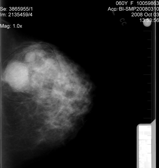
A patient without clinical symptoms had a mammogram in October 2008. The procedure caused intense persistent pain, swelling and development of a haematoma following mediolateral left breast compression. Three months later, a 9×11 cm mass developed within the same region. Core biopsies showed a necrotizing high-grade ductal carcinoma, with a high mitotic index. Owing to its extensive size, the patient began chemotherapy followed by trastuzumab and later radiotherapy to obtain clear margins for a subsequent mastectomy. The mastectomy in October 2009 revealed an inflammatory carcinoma, with 2 of 3 nodes infiltrated by the tumour. The stage IIIC tumour, oestrogen and progesterone receptor negative, was highly HER2 positive. A recurrence led to further chemotherapy in February 2011. In July 2011, another recurrence was removed from the mastectomy scar. She died of progressive disease in 2012. In this article, we discuss the potential influence of compression on the natural history of the tumour.
2016 BMJ Publishing Group Ltd.
References
-
- van Netten JP, Cann CA. Compression mammography and breast cancer: should pain be ignored? Cancer J 1996;9:278–9.
-
- van Netten JP, Cann SA, Hall JG. Mammographic controversies: time for informed consent? J Natl Cancer Inst 1997;89:1164–5. - PubMed
-
- Sapir R, Patlas M, Strano SD et al. Does mammography hurt? J Pain Symptoms Manage 2003;1:53–63. - PubMed
-
- Davey B. Pain during mammography: possible risk factors and ways to alleviate pain. Radiography 2007;3:229–34.
-
- Hugh TB. Cutaneous bruising after mammography. Med J Aust 1991;154:712. - PubMed
Publication types
MeSH terms
LinkOut - more resources
Full Text Sources
Other Literature Sources
Medical
Research Materials
Miscellaneous