. 2016 Nov;71(11):1030-1038.
doi: 10.1136/thoraxjnl-2015-207604. Epub 2016 Aug 31.

Hypoxia upregulates neutrophil degranulation and potential for tissue injury

Affiliations

Hypoxia upregulates neutrophil degranulation and potential for tissue injury

Kim Hoenderdos et al. Thorax. 2016 Nov.

Abstract

Background: The inflamed bronchial mucosal surface is a profoundly hypoxic environment. Neutrophilic airway inflammation and neutrophil-derived proteases have been linked to disease progression in conditions such as COPD and cystic fibrosis, but the effects of hypoxia on potentially harmful neutrophil functional responses such as degranulation are unknown.

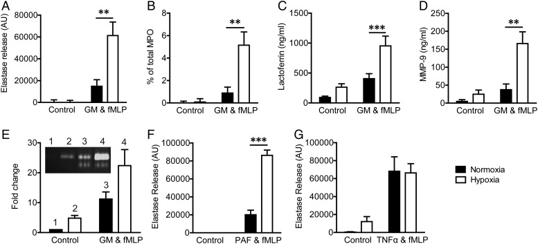
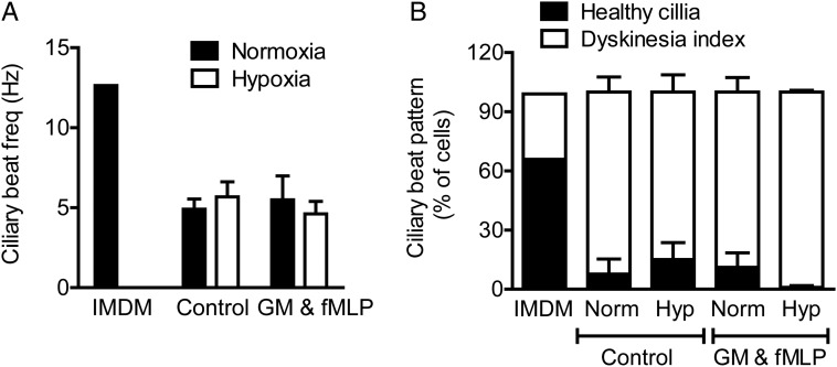
Methods and results: Following exposure to hypoxia (0.8% oxygen, 3 kPa for 4 h), neutrophils stimulated with inflammatory agonists (granulocyte-macrophage colony stimulating factor or platelet-activating factor and formylated peptide) displayed a markedly augmented (twofold to sixfold) release of azurophilic (neutrophil elastase, myeloperoxidase), specific (lactoferrin) and gelatinase (matrix metalloproteinase-9) granule contents. Neutrophil supernatants derived under hypoxic but not normoxic conditions induced extensive airway epithelial cell detachment and death, which was prevented by coincubation with the antiprotease α-1 antitrypsin; both normoxic and hypoxic supernatants impaired ciliary function. Surprisingly, the hypoxic upregulation of neutrophil degranulation was not dependent on hypoxia-inducible factor (HIF), nor was it fully reversed by inhibition of phospholipase C signalling. Hypoxia augmented the resting and cytokine-stimulated phosphorylation of AKT, and inhibition of phosphoinositide 3-kinase (PI3K)γ (but not other PI3K isoforms) prevented the hypoxic upregulation of neutrophil elastase release.

Conclusion: Hypoxia augments neutrophil degranulation and confers enhanced potential for damage to respiratory airway epithelial cells in a HIF-independent but PI3Kγ-dependent fashion.

Keywords: Airway Epithelium; COPD ÀÜ Mechanisms; Cystic Fibrosis; Innate Immunity; Neutrophil Biology; Respiratory Infection.


Conflict of interest statement

Conflicts of Interest: None declared.

Figures

Figure 1
Figure 1
Hypoxia augments neutrophil degranulation. (A–E) Hypoxia augments the release of all granule populations. Neutrophils were incubated under normoxia or hypoxia for 4 h, primed (granulocyte-macrophage colony stimulating factor (GM-CSF); 10 ng/mL, 30 min) or vehicle controlled and activated (formylated peptide (fMLP); 100 nM, 10 min), and the supernatants were assayed for the presence of granule proteins. Hypoxia augmented the release of active elastase (A, n=10, each performed in triplicate) as measured by elastase activity assay, active myeloperoxidase (MPO) (B, n=8, each performed in triplicate) as measured by the H2O2-dependent oxidation of O-dianisidine dihydrochloride, lactoferrin as measured by ELISA (C, n=6) and active matrix metalloproteinase-9 (MMP-9) as measured by ELISA (D, n=5, each performed in triplicate) and gelatine zymography (E, n=3, each performed in triplicate plus representative zymogram, inset). (F and G) Neutrophils were incubated under normoxia or hypoxia for 4 h prior to priming with platelet-activating factor (PAF) (1 µM, 5 min, n=4, each performed in triplicate) or TNFα (G, 20 ng/mL 30 min, n=10, each performed in triplicate) or media control and activation with fMLP (100 nM, 10 min), and the supernatants were assessed by elastase activity assay as above. Results represent mean±SEM. **p<0.01, ***p<0.005.
Figure 2
Figure 2
Hypoxia increases neutrophil supernatant-mediated epithelial injury. (A–C) Hypoxia increased the injurious potential of neutrophil supernatants to A549 cells. Confluent cell monolayers were exposed to diluted (1:2.5) supernatants (from neutrophils incubated under normoxic or hypoxic conditions for 4 h, without (control) or with GM-CSF (GM) and formylated peptide (fMLP) activation)±α1AT (46 μg/mL) for 48–72 h. At 48 h, cell layers were stained (A, representative images from n=6 experiments, each performed in triplicate) for cleaved caspase 3 (red), F-actin (green) and nuclei (blue). Survival of the A549 cells at 72 h was measured by MTT assay (B, n=11, each performed in triplicate; ***p<0.005, ****p<0.001). Cell layer detachment was quantified with ImageJ (C, n=3, three fields of view per well, performed in triplicate; *p<0.05, **p<0.01 (paired t test)). (D and E) Hypoxia increased the injurious potential of neutrophil supernatants to immortalised human bronchial epithelial cells28 (iHBECs). Confluent cell layers were exposed to neutrophil supernatants (1:2.5 dilution) for 48 h. At 48 h, cell layers were stained (D, representative images from n=3, each performed in triplicate) for cleaved caspase 3 (red), F-actin (green) and nuclei (blue). Cell layer detachment was quantified with ImageJ (E, n=3, each performed in triplicate). Results represent mean±SEM.
Figure 3
Figure 3
Neutrophil supernatants impair ciliary function. Supernatants from neutrophils incubated under normoxic or hypoxic conditions for 4 h, without (control) or with GM-CSF (GM) and formylated peptide (fMLP) activation were applied to strips of ciliated primary human bronchial epithelial cells for 2 h. Ciliary function was assessed by high-speed digital video microscopy allowing ciliary beat frequency (CBF, A) and detailed ciliary beat pattern (CBP, B) analysis as detailed in the Methods section. Results represent mean±SEM (n=3, three movies per ciliated strip of epithelial cells).
Figure 4
Figure 4
Hypoxic upregulation of neutrophil degranulation is not hypoxia-inducible factor (HIF)-dependent. (A and B) Hypoxia mimetics do not recapitulate the hypoxic upregulation of neutrophil degranulation. Neutrophils were incubated under normoxia for 4 h±DMOG (dimethyloxalyl glycine) (1 mM, A) or desferrioxamine (DFO) (1 mM, B) or were incubated under hypoxia (positive control), prior to priming with GM-CSF (GM) (10 ng/mL, 30 min) and activation with formylated peptide (fMLP) (100 nM, 10 min). Supernatants were assayed for active elastase. Results represent mean±SEM (n=5–6, each performed in triplicate). (C–E) Hypoxia does not increase granule protein transcription, translation or total protein. Neutrophils were incubated under normoxia or hypoxia for 4 h before RNA isolation or protein quantification. (C) Granule protein (NE, neutrophil elastase and MMP-9, matrix metalloproteinase-9) and BNIP (BCL2/adenovirus E1B 19 kd-interacting protein-2, positive control) gene expression as measured by quantitative PCR (qPCR); results represent mean±SEM (n=3, each performed in triplicate). (D) Gelatinase granule content measured by western blot analysis of MMP-9; results represent mean±SEM (n=3). (E) Hypoxic elastase release was not prevented when translation was inhibited by cycloheximide, present for the entire incubation, prior to stimulation with GM-CSF (GM) and fMLP. Results represent mean±SEM (n=5, each performed in triplicate). (F) Reoxygenation (15 min) of neutrophils before priming and activation does not significantly reduce degranulation as measured by the release of active elastase. Results represent mean±SEM (n=5, each performed in triplicate). *p<0.05, **p<0.01, ***p<0.005.
Figure 5
Figure 5
Hypoxic upregulation of neutrophil degranulation does not depend on cytoskeletal remodelling. (A) Neutrophils were incubated under normoxia or hypoxia for 4 h to activation with formylated peptide (fMLP) (100 nM, 5 min). Cells were stained for F-actin (green), neutrophil elastase (red) and nuclei (blue). Representative images are from n=3 experiments, each performed in triplicate. (B) Neutrophils were incubated under normoxia or hypoxia for 4 h prior to priming with cytochalasin B (Cyt B: 5 μg/mL, 5 min) and activation with fMLP (100 nM, 10 min); degranulation was measured by the release of active elastase. Results represent mean±SEM (n=10, each performed in triplicate). (C and D) Induction of actin polymerisation induces the formation of actin ‘rings’, but does not affect neutrophil degranulation. (C) Neutrophils incubated with jasplakinolide (1 μM, 5 min) prior to activation with fMLP (100 nM, 10 min). Cells were stained for F-actin (green) and nuclei (blue). (D) Neutrophils were incubated under normoxia or hypoxia for 4 h prior to priming with GM-CSF (GM) (10 ng/mL, 30 min), treatment with jasplakinolide (1 μM, 5 min) prior to activation with fMLP (100 nM, 10 min). Degranulation was measured by the release of active elastase. Results represent mean±SEM (n=4, each performed in triplicate). *p<0.05, ***p<0.005, ****p<0.001.
Figure 6
Figure 6
Phospholipase C (PLC) and calcium signalling regulate neutrophil degranulation but do not mediate the hypoxic uplift. Inhibition of the PLC pathway (A) or calcium signalling (B) abolished neutrophil elastase release induced by agonist stimulation under normoxia, and reduced but did not abolish degranulation under conditions of hypoxia. Increasing intracellular calcium (C) induced an increase in degranulation, but similarly did no abolish the hypoxic uplift induced by hypoxia. Neutrophils were incubated under normoxia or hypoxia for 4 h prior to priming with GM-CSF (GM)) (10 ng/mL, 30 min). Neutrophils were preincubated with a PLC inhibitor (U-73122, 2 μM) for 10 min prior to activation with formylated peptide (fMLP) (100 nM, 10 min). EGTA (2 mM) was added at the start of the normoxic/hypoxic incubation, and thapsigargin (100 nM) was added immediately prior to neutrophil activation with fMLP. Cells were spun down and degranulation measured by the release of active elastase. Results represent mean±SEM (n=4–5, each performed in triplicate). *p<0.05, **p<0.01, ****p<0.001.
Figure 7
Figure 7
Phosphoinositide 3-kinase (PI3K) signalling contributes to the hypoxic uplift of neutrophil degranulation. (A) Hypoxia induces neutrophil survival at 20 h, which is prevented by incubation with the pan-PI3K inhibitor LY294002 (10 μM). Results represent mean±SEM (n=4, each performed in triplicate). (B and C) Inhibition of the PI3K pathway inhibits degranulation and abrogates the hypoxic response. Neutrophils were incubated under normoxia or hypoxia for 4 h prior to priming with GM-CSF (GM) (10 ng/mL, 30 min). Cells were treated with a pan-PI3K inhibitor (LY294002, 10 μM), a PI3Kγ-selective inhibitor (AS605240, 3 μM), a PI3Kδ-selective inhibitor (IC87114, 3 μM), either for 10 min prior to activation with formylated peptide (fMLP) (B) or from the outset of the normoxic/hypoxic incubation (C). Degranulation was measured by the release of active elastase. Results represent mean±SEM (n=5). (D and E) Hypoxia increases AKT serine-473 phosphorylation after 15 min of GM-CSF stimulation in a PI3Kγ-dependent fashion. Neutrophils were incubated under normoxia or hypoxia for 4 h prior to priming with GM-CSF (10 ng/mL, 15 min). Isoform-selective PI3K inhibitors AS605240 (3 μM, PI3Kγ) and IC87114 (3 μM, PI3Kδ) were added at the start of the incubation. p-AKT was quantified by western blotting. Results represent mean±SEM (n=4–11). *p<0.05, **p<0.01.

Comment in

References

    1. Schmidt EP, Lee WL, Zemans RL, et al. . On, around, and through: neutrophil-endothelial interactions in innate immunity. Physiology (Bethesda) 2011;26:334–47. 10.1152/physiol.00011.2011 - DOI - PMC - PubMed
    1. Abraham E, Carmody A, Shenkar R, et al. . Neutrophils as early immunologic effectors in hemorrhage- or endotoxemia-induced acute lung injury. Am J Physiol 2000;279:L1137–45. - PubMed
    1. Brazil JC, Louis NA, Parkos CA. The role of polymorphonuclear leukocyte trafficking in the perpetuation of inflammation during inflammatory bowel disease. Inflamm Bowel Dis 2013;19:1556–65. 10.1097/MIB.0b013e318281f54e - DOI - PMC - PubMed
    1. Datta S, Kundu S, Ghosh P, et al. . Correlation of oxidant status with oxidative tissue damage in patients with rheumatoid arthritis. Clin Rheumatol 2014;33:1557–64. 10.1007/s10067-014-2597-z - DOI - PubMed
    1. Worthen GS, Haslett C, Rees AJ, et al. . Neutrophil-mediated pulmonary vascular injury. Synergistic effect of trace amounts of lipopolysaccharide and neutrophil stimuli on vascular permeability and neutrophil sequestration in the lung. Am Rev Respir Dis 1987;136:19–28. - PubMed

Publication types

MeSH terms