

Effects of Alpha-Synuclein on Primary Spinal Cord Neurons Associated with Apoptosis and CNTF Expression
- PMID: 27581683
- PMCID: PMC11482142
- DOI: 10.1007/s10571-016-0420-x
Effects of Alpha-Synuclein on Primary Spinal Cord Neurons Associated with Apoptosis and CNTF Expression
Abstract
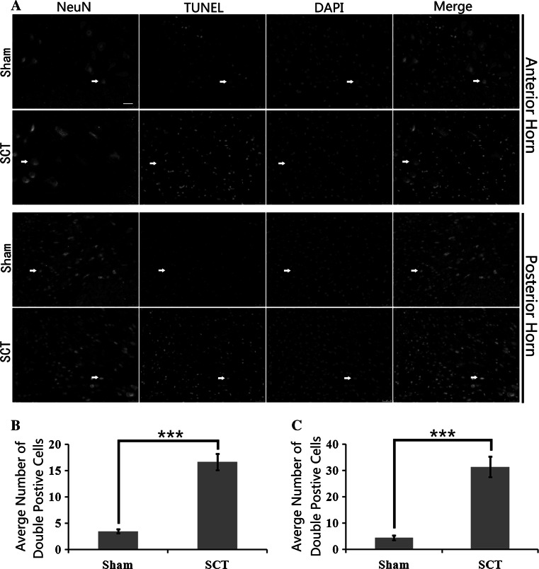
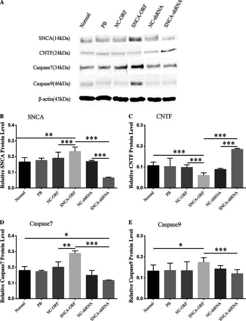
Spinal cord injury (SCI) often causes neurological deficits with poor recovery; the treatment, however, is far from satisfaction, and the mechanisms remain unclear. Using immunohistochemistry and western blotting analysis, we found α-synuclein (SNCA) was significantly up-regulated in the spinal caudal segment of rats subjected to spinal cord transection at 3 days post-operation. Moreover, the role of SNCA on neuronal growth and apoptosis in vitro was determined by using overexpressing and interfering SNCA recombined plasmid vectors, and the underlying mechanism was detected by QRT-PCR and western blotting. Spinal neurons transfected with SNCA-shRNA lentivirus gave rise to an optimal neuronal survival, while it results in cell apoptosis in SNCA-ORF group. In molecular level, SNCA silence induced the up-regulation of CNTF and down-regulation of Caspase7/9. Together, endogenous SNCA plays a crucial role in spinal neuronal survival, in which the underlying mechanism may be linked to the regulation both apoptotic genes (Caspase7/9) and CNTF. The present findings therefore provide novel insights into the role of SNCA in spinal cord and associated mechanism, which may provide novel cue for the treatment of SCI in future clinic trials.
Keywords: Alpha-synuclein; Apoptotic gene; Ciliary neurotrophic factor; Primary spinal neurons; Spinal cord transection.
Conflict of interest statement
All authors declare that they have no conflict of interest.
Figures


Similar articles
-
Lentivirus-mediated downregulation of α-synuclein reduces neuroinflammation and promotes functional recovery in rats with spinal cord injury.J Neuroinflammation. 2019 Dec 30;16(1):283. doi: 10.1186/s12974-019-1658-2. J Neuroinflammation. 2019. PMID: 31888724 Free PMC article.
-
Knockdown of α-synuclein in cerebral cortex improves neural behavior associated with apoptotic inhibition and neurotrophin expression in spinal cord transected rats.Apoptosis. 2016 Apr;21(4):404-20. doi: 10.1007/s10495-016-1218-5. Apoptosis. 2016. PMID: 26822976
-
Changes in expression of ciliary neurotrophic factor (CNTF) and CNTF-receptor alpha after spinal cord injury.J Neurobiol. 1997 Mar;32(3):251-61. J Neurobiol. 1997. PMID: 9058319
-
Chronically increased ciliary neurotrophic factor and fibroblast growth factor-2 expression after spinal contusion in rats.J Comp Neurol. 2008 Sep 10;510(2):129-44. doi: 10.1002/cne.21787. J Comp Neurol. 2008. PMID: 18615534 Free PMC article.
-
Neurotrophic factors for spinal cord repair: Which, where, how and when to apply, and for what period of time?Brain Res. 2015 Sep 4;1619:36-71. doi: 10.1016/j.brainres.2014.10.049. Epub 2014 Nov 1. Brain Res. 2015. PMID: 25451132 Review.
Cited by
-
Transcriptomic analysis of α-synuclein knockdown after T3 spinal cord injury in rats.BMC Genomics. 2019 Nov 14;20(1):851. doi: 10.1186/s12864-019-6244-6. BMC Genomics. 2019. PMID: 31726970 Free PMC article.
-
Lentivirus-mediated downregulation of α-synuclein reduces neuroinflammation and promotes functional recovery in rats with spinal cord injury.J Neuroinflammation. 2019 Dec 30;16(1):283. doi: 10.1186/s12974-019-1658-2. J Neuroinflammation. 2019. PMID: 31888724 Free PMC article.
-
α-Synuclein in traumatic and vascular diseases of the central nervous system.Aging (Albany NY). 2020 Nov 7;12(21):22313-22334. doi: 10.18632/aging.103675. Epub 2020 Nov 7. Aging (Albany NY). 2020. PMID: 33188159 Free PMC article. Review.
-
Synucleinopathy in Amyotrophic Lateral Sclerosis: A Potential Avenue for Antisense Therapeutics?Int J Mol Sci. 2022 Aug 19;23(16):9364. doi: 10.3390/ijms23169364. Int J Mol Sci. 2022. PMID: 36012622 Free PMC article. Review.
-
Protein Network Analysis of the Serum and Their Functional Implication in Patients Subjected to Traumatic Brain Injury.Front Neurosci. 2019 Jan 31;12:1049. doi: 10.3389/fnins.2018.01049. eCollection 2018. Front Neurosci. 2019. PMID: 30766469 Free PMC article.
References
-
- Abbaszadeh HA, Tiraihi T, Noori-Zadeh A, Delshad AR, Sadeghizade M, Taheri T (2015) Human ciliary neurotrophic factor-overexpressing stable bone marrow stromal cells in the treatment of a rat model of traumatic spinal cord injury. Cytotherapy 17:912–921 - PubMed
-
- Beattie MS, Hermann GE, Rogers RC, Bresnahan JC (2002) Cell death in models of spinal cord injury. Prog Brain Res 137:37–47 - PubMed
-
- Bregman BS, McAtee M, Dai HN, Kuhn PL (1997) Neurotrophic factors increase axonal growth after spinal cord injury and transplantation in the adult rat. Exp Neurol 148:475–494 - PubMed
MeSH terms
Substances
LinkOut - more resources
Full Text Sources
Other Literature Sources
Miscellaneous
