. 2016 Oct 14;90(21):10022-10038.
doi: 10.1128/JVI.01196-16. Print 2016 Nov 1.

Packaging and Prefusion Stabilization Separately and Additively Increase the Quantity and Quality of Respiratory Syncytial Virus (RSV)-Neutralizing Antibodies Induced by an RSV Fusion Protein Expressed by a Parainfluenza Virus Vector

Affiliations

Packaging and Prefusion Stabilization Separately and Additively Increase the Quantity and Quality of Respiratory Syncytial Virus (RSV)-Neutralizing Antibodies Induced by an RSV Fusion Protein Expressed by a Parainfluenza Virus Vector

Bo Liang et al. J Virol. .

Abstract

Human respiratory syncytial virus (RSV) and human parainfluenza virus type 3 (HPIV3) are major pediatric respiratory pathogens that lack vaccines. A chimeric bovine/human PIV3 (rB/HPIV3) virus expressing the unmodified, wild-type (wt) RSV fusion (F) protein from an added gene was previously evaluated in seronegative children as a bivalent intranasal RSV/HPIV3 vaccine, and it was well tolerated but insufficiently immunogenic for RSV F. We recently showed that rB/HPIV3 expressing a partially stabilized prefusion form (pre-F) of RSV F efficiently induced "high-quality" RSV-neutralizing antibodies, defined as antibodies that neutralize RSV in vitro without added complement (B. Liang et al., J Virol 89:9499-9510, 2015, doi:10.1128/JVI.01373-15). In the present study, we modified RSV F by replacing its cytoplasmic tail (CT) domain or its CT and transmembrane (TM) domains (TMCT) with counterparts from BPIV3 F, with or without pre-F stabilization. This resulted in RSV F being packaged in the rB/HPIV3 particle with an efficiency similar to that of RSV particles. Enhanced packaging was substantially attenuating in hamsters (10- to 100-fold) and rhesus monkeys (100- to 1,000-fold). Nonetheless, TMCT-directed packaging substantially increased the titers of high-quality RSV-neutralizing serum antibodies in hamsters. In rhesus monkeys, a strongly additive immunogenic effect of packaging and pre-F stabilization was observed, as demonstrated by 8- and 30-fold increases of RSV-neutralizing serum antibody titers in the presence and absence of added complement, respectively, compared to pre-F stabilization alone. Analysis of vaccine-induced F-specific antibodies by binding assays indicated that packaging conferred substantial stabilization of RSV F in the pre-F conformation. This provides an improved version of this well-tolerated RSV/HPIV3 vaccine candidate, with potently improved immunogenicity, which can be returned to clinical trials.

Importance: Human respiratory syncytial virus (RSV) and human parainfluenza virus type 3 (HPIV3) are major viral agents of acute pediatric bronchiolitis and pneumonia worldwide that lack vaccines. A bivalent intranasal RSV/HPIV3 vaccine candidate consisting of a chimeric bovine/human PIV3 (rB/HPIV3) strain expressing the RSV fusion (F) protein was previously shown to be well tolerated by seronegative children but was insufficiently immunogenic for RSV F. In the present study, the RSV F protein was engineered to be packaged efficiently into vaccine virus particles. This resulted in a significantly enhanced quantity and quality of RSV-neutralizing antibodies in hamsters and nonhuman primates. In nonhuman primates, this effect was strongly additive to the previously described stabilization of the prefusion conformation of the F protein. The improved immunogenicity of RSV F by packaging appeared to involve prefusion stabilization. These findings provide a potently more immunogenic version of this well-tolerated vaccine candidate and should be applicable to other vectored vaccines.


Figures

FIG 1
FIG 1
rB/HPIV3 vectors expressing versions of the RSV F protein modified to contain CT and TMCT domains from the vector BPIV3 F protein. (A) Gene inserts expressing the indicated versions of RSV F were cloned into the second gene position (between the N and P genes) in rB/HPIV3 as previously described (28). HEK/opt encodes a wt RSV F protein (dark blue) (29) containing HEK assignments 66E and 101P (indicated by asterisks; see Results for an explanation) and was expressed from a codon-optimized ORF (GeneArt) (29). B3CT and B3TMCT are derivatives of HEK/opt modified to contain the CT and TMCT domains of BPIV3 F (red). Constructs with the designation DS contain an introduced disulfide bond (S155C and S290C mutations; indicated by stars) that stabilizes RSV in the pre-F conformation (29). (B) The C-terminal sequences of BPIV3 F and the B3CT and B3TMCT forms of F are aligned to that of wt RSV F (42, 53, 54) to indicate their predicted TM and CT domains, based on hydrophobicity and sequence content.
FIG 2
FIG 2
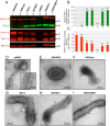
Packaging of RSV F into rB/HPIV3 virion particles. (A to C) Sucrose gradient-purified preparations of the indicated rB/HPIV3 vectors or wt RSV (0.5 μg of protein per sample) were analyzed by Western blotting using antibodies specific to RSV F, HPIV3 HN, HPIV3 F, and BPIV3 N, as described in Materials and Methods. (A) Western blot images. (B) Quantification of RSV F packaging efficiency, based on the ratio of RSV F to BPIV3 N, calculated from the data in panel A. All ratios were normalized against that of HEK/opt, which was set at 1. Packaging efficiency values are indicated at the top of the bars. The packaging efficiency of wt RSV F was not calculated (NA) because of the lack of a BPIV3 N value. (C) Quantification of HPIV3 F packaging efficiency. The ratio of total HPIV3 F (F0 + F1) to BPIV3 N was calculated and normalized against that of the empty rB/HPIV3 vector, which was set at 1. Packaging efficiency values are indicated at the top of the bars. (D to I) Immunoelectron microscopy analysis of RSV F in rB/HPIV3 virions. Sucrose gradient-purified virus preparations of the indicated rB/HPIV3 vectors and wt RSV were fixed with paraformaldehyde and incubated with an RSV F-specific mouse MAb, followed by a goat anti-mouse IgG specific secondary antibody conjugated with 5-nm-diameter gold particles, and subjected to transmission electron microscopy. A representative virion image for each virus is shown, and arrows indicate representative virion-associated gold particles. (D) Filamentous wt RSV particle exhibiting abundant gold particles; (E) spherical empty rB/HPIV3 vector particle that remained unlabeled; (F) HEK/opt virion with only trace amounts of gold particles; (G to I) B3CT, B3TMCT, and DS/B3TMCT virions, respectively, showing abundant labeling with gold particles. The RSV F protein detected on the latter vector particles (G to I) had an abundance and distribution pattern similar to those of wt RSV (D).
FIG 3
FIG 3
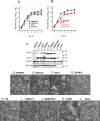
Replication, intracellular protein expression, and syncytium formation in Vero cells by rB/HPIV3 vectors expressing various forms of RSV F. (A and B) Multicycle replication of rB/HPIV3 vectors in Vero cells at 32°C. Vero cells were infected with the following vectors at an MOI of 0.01 TCID50 per cell, in triplicate: empty rB/HPIV3, HEK/opt, B3CT, and B3TMCT (A) or empty rB/HPIV3, DS, DS/B3CT, and DS/B3TMCT (B). A portion of the medium (0.5 ml out of 2 ml) was collected from the infected cell culture and replaced with fresh medium every 24 h during a period of 6 days. Viral titers in medium supernatants were determined by TCID50 assays in LLC-MK2 cells at 32°C. (C) Western blot analysis of the expression of RSV F by vectors and wt RSV in Vero cells at 32°C. Vero cells were infected at an MOI of 10 TCID50 per cell with the indicated vectors or 3 PFU per cell with wt RSV and then incubated at 32°C. Cell lysates were harvested at 48 h p.i., and expression of RSV F was analyzed by Western blotting. Analysis of BPIV3 N and GAPDH provided controls for vector expression and gel loading, respectively. Cleaved (F1) and uncleaved (F0) forms of RSV F are indicated. (D to L) Syncytium formation in Vero cell monolayers infected with empty rB/HPIV3 (D), HEK/opt (E), B3CT (F), B3TMCT (G), DS (H), DS/B3CT (I), or DS/B3TMCT (J), at an MOI of 10 TCID50 per cell, or with wt RSV (K) at an MOI of 3 PFU per cell, or mock infected (L). Images were acquired at 48 h p.i.
FIG 4
FIG 4
Levels of serum neutralizing antibodies induced by rB/HPIV3 vectors and wt RSV in hamsters. Hamsters (n = 6) were inoculated intranasally with 105 TCID50 of rB/HPIV3 vectors and 106 PFU of wt RSV (A2) in a 0.1-ml inoculum. Serum samples were collected at day 28 postinoculation. RSV-neutralizing antibody titers were determined by a 60% plaque reduction neutralization test (PRNT60) with (A) or without (B) guinea pig complement. (C) HPIV3-neutralizing antibody titers were determined by PRNT60 with complement. The detection limit of each assay is indicated by a dotted line. The colored symbols represent individual animal titers. The mean titer for each group is shown as a vertical bar, with error bars representing standard errors of the means (SEMs), and the value of the mean titer is shown above the bar. Mean titers were assigned to different groups (A, B, C, and D) by the Tukey-Kramer test. Mean titers with different letters are statistically different (P < 0.05). Titers with two letters are not significantly different from those with either letter.
FIG 5
FIG 5
Protection of immunized hamsters against wt RSV challenge. Hamsters (n = 6 per virus) that were immunized with the indicated vectors or wt RSV (from the results shown in Table 2) were challenged i.n. on day 30 postimmunization with 106 PFU of wt RSV in a 0.1-ml inoculum. At 3 days postchallenge, hamsters were sacrificed, and nasal turbinates (A) and lungs (B) were collected. Tissue homogenates were prepared, and virus titers were determined by plaque assay on Vero cells at 37°C. Each symbol represents the RSV titer of an individual animal. Mean viral titers for the groups are shown as horizontal lines, with the numerical values indicated, and the statistical significance of differences between mean titers was determined by one-way ANOVA with the Tukey-Kramer test, using Prism software, and is indicated as follows: ns, not significant; *, P ≤ 0.05; **, P ≤ 0.01; ***, P ≤ 0.001; and ****, P ≤ 0.0001. The detection limit of the assay was 2.7 log10 PFU/g of tissue and is indicated with a dotted line. The data for rB/HPIV3, HEK/opt, DS, and wt RSV were published previously (29) and were included in this figure for comparison.
FIG 6
FIG 6
Replication of rB/HPIV3 vectors in the respiratory tract of rhesus macaques. Rhesus monkeys were infected by the combined i.n. and i.t. routes with the indicated rB/HPIV3 vectors at a dose of 106 TCID50 in a 1-ml inoculum per route. Groups of five, five, and four monkeys per group were infected with non-HEK/non-opt, DS, and DS/B3TMCT, respectively. Nasopharyngeal swabs and tracheal lavage fluids were collected on the indicated days. Viral titers in collected samples were determined by TCID50 assay on MK2 cells at 32°C. The mean titers for the groups at each time point are plotted, and error bars represent SEMs.
FIG 7
FIG 7
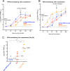
RSV- and HPIV3-neutralizing serum antibodies induced in rhesus macaques immunized with rB/HPIV3 vectors expressing RSV F. Sera were collected from the immunized monkeys at 0, 14, 21, and 28 days as part of the experiment described for Fig. 6. At 28 days postimmunization, all monkeys were challenged by the combined i.n. and i.t. routes with 106 PFU of wt RSV in a 1-ml inoculum per site, and sera were collected on days 35 and 56. (A) Mean titers of HPIV3-neutralizing serum antibodies determined with HPIV3 PRNT60 in the presence of complement. (B) Mean titers of RSV-neutralizing serum antibodies determined with RSV PRNT60 in the presence of complement. (C) Titers of RSV-neutralizing serum antibodies at day 28 postimmunization determined by RSV PRNT60 in the absence of complement. Each symbol represents an individual rhesus monkey, and the mean titer for each group is indicated as a solid horizontal line, with the mean value shown beside the line. For all panels, the statistical significance of differences in mean PRNT60 values between pairs of groups at each time point was determined by the pairwise Student t test, and P values are indicated by asterisks (**, P ≤ 0.01; and ***, P ≤ 0.001). The detection limit of each assay is shown with a dotted horizontal line.
FIG 8
FIG 8
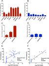
Pre-F-specific serum antibodies induced in hamsters and rhesus monkeys by rB/HPIV3 vectors and their correlation with titers of high-quality RSV-neutralizing antibodies. Hamster and rhesus monkey sera from the experiments performed for Fig. 4 and 7 were assayed by biolayer interferometry (BLI) for the ability to bind to the biosensor-tagged pre-F or post-F protein in the presence or absence of competing soluble post-F protein. Binding activity was calculated as follows: (binding in the presence of competing post-F)/(binding in the absence of competing post-F) × 100. (A and C) Percentages of pre-F-specific antibody binding retained in the presence of competing post-F in hamster sera (A) and rhesus monkey sera (C). (B and D) Percentages of post-F-binding antibodies retained in the presence of competing post-F in hamster sera (B) and rhesus monkey sera (D). Note that the “postfusion” construct included in panels A and B is an rB/HPIV3 vector expressing post-F that was not shown in Fig. 4 but was part of the same experiment and was described previously (29). (E and F) Correlations of the percentages of pre-F-binding antibodies from panels A and C with RSV-neutralizing antibody titers, assayed in the absence of complement, in hamster sera (E) and rhesus monkey sera (F) from the experiments performed for Fig. 4 and 7. The fitting line was generated with a linear regression model. Pearson's correlation coefficient (Pearson R), the sample size (N), and the P value are indicated on the graph. The sera used for panels A, B, and E comprised 45 of the 48 specimens representing the vectors in Fig. 4A and B; three sera (two for B3CT and one for the postfusion construct) were omitted due to low pre-F-binding titers.

References

    1. Nair H, Nokes DJ, Gessner BD, Dherani M, Madhi SA, Singleton RJ, O'Brien KL, Roca A, Wright PF, Bruce N, Chandran A, Theodoratou E, Sutanto A, Sedyaningsih ER, Ngama M, Munywoki PK, Kartasasmita C, Simoes EA, Rudan I, Weber MW, Campbell H. 2010. Global burden of acute lower respiratory infections due to respiratory syncytial virus in young children: a systematic review and meta-analysis. Lancet 375:1545–1555. doi:10.1016/S0140-6736(10)60206-1. - DOI - PMC - PubMed
    1. Murphy BR, Prince GA, Collins PL, Van Wyke Coelingh K, Olmsted RA, Spriggs MK, Parrott RH, Kim HW, Brandt CD, Chanock RM. 1988. Current approaches to the development of vaccines effective against parainfluenza and respiratory syncytial viruses. Virus Res 11:1–15. - PubMed
    1. Klein MI, Coviello S, Bauer G, Benitez A, Serra ME, Schiatti MP, Delgado MF, Melendi GA, Novalli L, Pena HG, Karron RA, Kleeberger SR, Polack FP. 2006. The impact of infection with human metapneumovirus and other respiratory viruses in young infants and children at high risk for severe pulmonary disease. J Infect Dis 193:1544–1551. doi:10.1086/503806. - DOI - PubMed
    1. Ottolini MG, Porter DD, Hemming VG, Prince GA. 2000. Enhanced pulmonary pathology in cotton rats upon challenge after immunization with inactivated parainfluenza virus 3 vaccines. Viral Immunol 13:231–236. doi:10.1089/vim.2000.13.231. - DOI - PubMed
    1. Kim HW, Canchola JG, Brandt CD, Pyles G, Chanock RM, Jensen K, Parrott RH. 1969. Respiratory syncytial virus disease in infants despite prior administration of antigenic inactivated vaccine. Am J Epidemiol 89:422–434. - PubMed

MeSH terms

Substances