. 2016 Aug 31;16(1):30.
doi: 10.1186/s12861-016-0131-z.

Impairment of Wnt11 function leads to kidney tubular abnormalities and secondary glomerular cystogenesis

Affiliations

Impairment of Wnt11 function leads to kidney tubular abnormalities and secondary glomerular cystogenesis

Irina I Nagy et al. BMC Dev Biol. .

Abstract

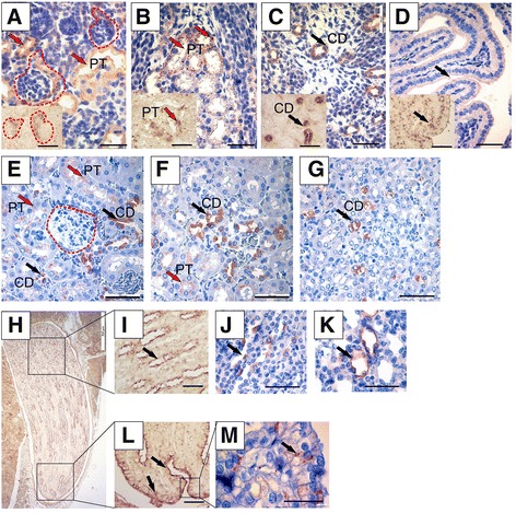
Background: Wnt11 is a member of the Wnt family of secreted signals controlling the early steps in ureteric bud (UB) branching. Due to the reported lethality of Wnt11 knockout embryos in utero, its role in later mammalian kidney organogenesis remains open. The presence of Wnt11 in the emerging tubular system suggests that it may have certain roles later in the development of the epithelial ductal system.

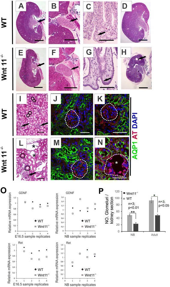
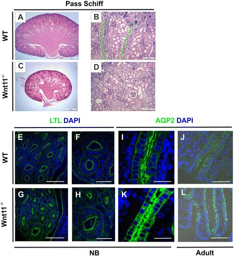
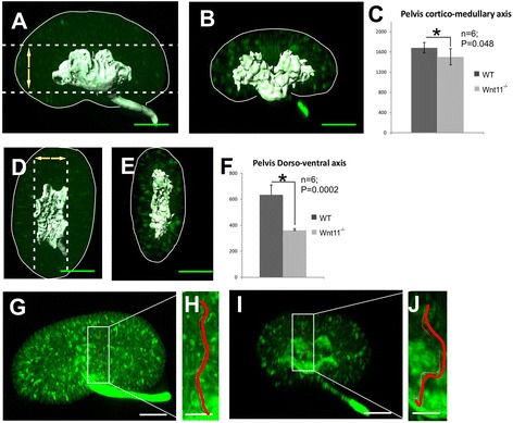
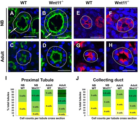
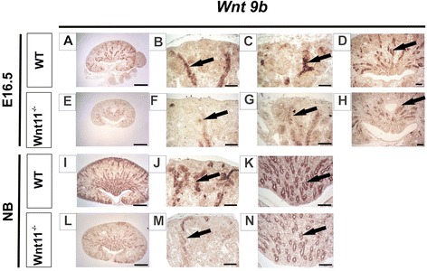
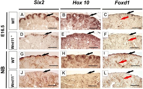
Results: The Wnt11 knockout allele was backcrossed with the C57Bl6 strain for several generations to address possible differences in penetrance of the kidney phenotypes. Strikingly, around one third of the null mice with this inbred background survived to the postnatal stages. Many of them also reached adulthood, but urine and plasma analyses pointed out to compromised kidney function. Consistent with these data the tubules of the C57Bl6 Wnt11 (-/-) mice appeared to be enlarged, and the optical projection tomography indicated changes in tubular convolution. Moreover, the C57Bl6 Wnt11 (-/-) mice developed secondary glomerular cysts not observed in the controls. The failure of Wnt11 signaling reduced the expression of several genes implicated in kidney development, such as Wnt9b, Six2, Foxd1 and Hox10. Also Dvl2, an important PCP pathway component, was downregulated by more than 90 % due to Wnt11 deficiency in both the E16.5 and NB kidneys. Since all these genes take part in the control of UB, nephron and stromal progenitor cell differentiation, their disrupted expression may contribute to the observed anomalies in the kidney tubular system caused by Wnt11 deficiency.

Conclusions: The Wnt11 signal has roles at the later stages of kidney development, namely in coordinating the development of the tubular system. The C57Bl6 Wnt11 (-/-) mouse generated here provides a model for studying the mechanisms behind tubular anomalies and glomerular cyst formation.

Keywords: Epithelial mesenchyme tissue interactions; Glomerular cysts; Tubule morphogenesis; Wnt signaling.


Figures

Fig. 1
Fig. 1
Wnt11 is expressed in the cells of the cortical, medullary and papilla tubules, including the Bellini ducts of the terminal papilla. Kidneys from NB (ad) and adult (em) mice were prepared and sectioned for in situ hybridization (ad inserts, h, i, l) or immunohistochemistry (ad large images, eg, j, k, m). Counterstaining was performed with haematoxylin (ad large images, eg, j, k, m). ad In the NB kidney, Wnt11 is expressed in the maturing proximal tubules (PT) (a, red arrows), the more mature proximal tubules (b and insert, red arrows), the collecting duct (CD) tubules (c and insert, black arrows) and sporadically in the terminal papillary tubules (d and insert, black arrows). eg Wnt11-positive cells in the cortical (e) and medullary (f, g) areas of the adult kidney. Expression is sporadic in the proximal tubule cells (e, f, red arrows) and more intense in the collecting duct cells (eg, black arrows). hm) The Wnt11 gene and protein detected in the papilla of the adult mouse kidney is located in the papillary duct tubular cells (i, j, k, arrows) and Bellini ducts cells (l, m, arrows). Wnt11 is not expressed in the glomerular tuft (a, e, areas circled by red dotted line). Bars: ag and im, 50 μm; h, 500 μm
Fig. 2
Fig. 2
Wnt11 −/− in C57Bl6 mice leads to kidney hypoplasia, abnormal morphology of the papilla and Bellini ducts, and glomerular cysts formation. The kidneys were dissected from 4-5-months-old C57Bl6 Wnt11 −/− and Wnt11 +/+ (WT) mice and sectioned rostro-caudally. a, d The WT kidney has a normal papilla morphology (arrow). b A higher magnification of WT papilla sections. c A high-power illustration of the epithelial cells in the region of the terminal papilla, showing Bellini duct cells with a columnar morphology (arrow). eg Wnt11deficiency in the C57Bl6 mouse leads to a hypoplastic kidney (compare e with a), with a typical abnormal appearance of the kidney papilla cells (compare e, f with a, b, arrows) and altered morphology of the Bellini duct cells towards a more squamous appearance (compare g with c, arrows). h An example of a very severe hypoplastic kidney in an adult C57Bl6 Wnt11 −/− mouse with large cysts (arrow). ik A WT kidney also depicting tubular cross-sections (i, circled areas) and glomeruli (i, arrow and j, k, dotted circled area). ln Typical findings in C57Bl6 Wnt11 −/− kidneys are cortical glomerular cysts in a rudimentary glomerular tuft (l, n, stars), enlarged proximal tubules (l, circled areas) and larger glomeruli (l, arrow). m, n Immunohistochemistry of proximal tubule marker Aquaporin 1 (AQP1) in green and glomerular podocytes cytoskeleton marker acetylated tubulin (AT) in red, nuclei marked with DAPI (blue). Note that the Wnt11 deficiency changes the Bowman capsule and the podocytes. No significant difference was noticed between the Wnt11 −/− glomeruli and control glomeruli prior to cyst development (compare m with j, area circled by the dotted line). Note the intense AT staining in the glomerular rudiments of the Wnt11 −/− kidneys (compare n with k, circled areas). o Relative mRNA levels of GDNF and Ret in Wnt11 −/− and WT kidneys examined by qRT-PCR. The data were normalized to GAPDH expression. p Number of glomeruli per kidney section in Wnt11 −/− and WT kidneys. ah; i, l Masson Trichrome staining. Bars: a, d, e, h 2 mm; b, f 500 μm; i, l 100 μm; c, g, j, k, m, n 50 μm. *P < 0.05, **P < 0.01
Fig. 3
Fig. 3
Wnt11 signaling is coupled to the development of the collecting duct and the kidney medullary tubular architecture. Adult WT and Wnt11 −/− kidneys were prepared, sectioned and processed for immunohistochemistry with the collecting duct (CD) marker Aquaporin 2 (AQP2), the PT and loop of Henle marker lectin LTL, and DAPI, depicting the nucleus. Periodic Acid Schiff (PAS) staining was used for the histological examination. ad The control kidney architecture, with longitudinal medullary rays consisting of the collecting ducts (green line), is deregulated in the Wnt11 −/− mice (compare c, d with a, b). el Confocal microscopy images with phase contrast. Note the control morphology of the terminal papillary tubules and Bellini ducts (e, f) and the narrower cross-section of the Bellini ducts in the Wnt11 −/− papilla (compare g, h with e, f). The high power micrographs depict AQP2 accumulation on the luminal side of the terminal papillary tubules in the WT (i, j), whereas AQP2 is also present on the basolateral side of the Wnt11 −/− cells (k, l). The papilla epithelial cells at the tip region have adopted an appearance similar to squamous cells (compare k, l with i, j). ah, i, k) NB. j, l Adult. Bars: a, c 500 μm; b, d, e, g 100 μm; f, h, j, l 50 μm; i, k 25 μm
Fig. 4
Fig. 4
Optical projection tomography shows Wnt11 dependent changes in the three-dimensional structure of the kidney. Kidneys of NB mice were dissected from the litters of Wnt11 +/− mating’s, processed and stained as whole-mount specimens with cytokeratin-Troma-1 antibody, and then subjected to optical projection tomography (OPT). The OPT data were analysed with 3D Imaris and Drishti software to examine the influence of Wnt11 knockout on the 3D layout of the kidney and its tubules. Troma-1 immunohistochemistry enables 3D presentation of the tubular projections in the kidney. Measurements of the Wnt11 −/− and WT kidney pelvis axes in the cortical-medullary/rostro-caudal (ac) and dorsal-ventral axis (df) views. Wnt11 −/− kidney tubule reconstruction pointed to changes in the overall tubule convolution as compared with the controls. gj One cortical-medullary tubule projection is highlighted in red in j, compare with (h). Bars: a, b, d, e, g, i 80 μm; h, j 300 μm
Fig. 5
Fig. 5
Wnt11 deficiency leads to the heterogeneity in cell numbers in specific tubular segmental regions. Kidneys from WT and Wnt11 −/− mice were prepared, sectioned and processed for immunohistochemistry with markers lectin LTL for the proximal tubule, in green, AQP2 for the collecting ducts, in red, and DAPI, for nuclei, in blue. ah The staining of the cross-sections of the tubules at corresponding spatial location emphasizes that Wnt11 deficiency leads to enlarged luminal diameter of the proximal tubules and collecting ducts in the kidney of NB (compare b, f with a, e, circled area) and adult mice (compare d, h with c, g, circled area). Note that the brush border, depicted with the LTL lectin marker, is more prominent and also abnormally organized in the proximal tubular cells of the Wnt11 −/− kidneys relative to the controls (compare b with a, arrows). i, j Histograms showing estimated percentages of the tubules having the given numbers of cells per cross-section. The Wnt11 deficient proximal and collecting ducts had increased percentages of tubules with an abnormally high cell number per cross-section relative to the values obtained from WT kidneys. Each colour indicates the number of cells/tubule cross-section (from 3 to 9–10 cells). Note that this phenotype was more obvious in the kidneys of the adult Wnt11-deficient mice. Bars: 25 μm
Fig. 6
Fig. 6
Wnt11 deficiency reduces cell proliferation and increases apoptosis in the kidney. Kidneys were prepared from WT and Wnt11 −/− mice and processed for immunohistochemistry. The sections were stained with phospho-Histone-3 (P-H3) antibodies, LTL and DAPI for cell proliferation and TUNEL for apoptosis. ad Representative cell proliferation images obtained by confocal microscopy (a, c). Corresponding brightfield images are also shown (b, d). Wnt11 −/− reduced cell proliferation in the nephron-forming cortex of C57Bl6 mice, as judged by the presence of P-H3 staining (in red) mainly in the non-LTL+ cells (green) (compare cd with ab, several proliferating cells marked with arrows). eh Cells in the developing cortex region underwent increased apoptosis in the absence of Wnt11 signaling, as indicated by the TUNEL assay (compare gh with ef, several apoptotic cells marked with arrows). i Quantification of TUNEL data (percentage of apoptotic cells in Wnt11 −/− and control samples) and P-H3 data (percentage of proliferating cells in Wnt11 −/− and control samples). Bars: 50 μm
Fig. 7
Fig. 7
Wnt9b, a UB and nephron regulator signal, is reduced in Wnt11 −/− kidneys. Kidneys were prepared from WT and Wnt11 −/− mice, sectioned and subjected to in situ hybridization with the Wnt9b probe. ah Wnt9b, which is expressed in the UB and the collecting duct (CD) cells derived from it, was notably reduced in the case of Wnt11 knockout, especially in the CD cells situated in the subcortical region (compare fg with bc, arrows). Note the decreased expression of Wnt9b in the medullary CD as well (compare h with d). in Wnt9bremains reduced in the CD of the NB mouse kidney (compare mn with jk, arrows). Bars: a, e, i, l 500 μm; b, c, f, g 50 μm, d, h, j, k, m, n 100 μm
Fig. 8
Fig. 8
Failure in Wnt11 function has an impact on the progenitors of the nephrons and the kidney stroma. Kidneys were prepared from WT and Wnt11-deficient mice, sectioned and subjected to in situ hybridization with the Six2, Hox10 and Foxd1 probes, depicting the nephron and the stromal progenitors, respectively. Six2, which is expressed in the nephron-forming progenitor cells, was severely reduced in the absence of Wnt11 (compare d, j with a, g, arrows). Similarly, Hox10 and Foxd1, which are expressed in the precursors of the renal stroma, became deregulated at E16.5 and were reduced the neonatal mice (compare b, c, h, i with e, f, k, l, black arrows). Note that the Foxd1 gene continues to be expressed in cells located deeper in the developing kidney (compare f, l with c, i, red arrows). The image exposure time was equivalent between all the samples of the same developmental stage for a given probe. Bar: al, 100 μm

Similar articles

Cited by

References

    1. Halt K, Vainio S. Coordination of kidney organogenesis by Wnt signaling. Pediatr Nephrol. 2014;29(4):737–744. doi: 10.1007/s00467-013-2733-z. - DOI - PMC - PubMed
    1. Majumdar A, Vainio S, Kispert A, McMahon J, McMahon AP. Wnt11 and Ret/Gdnf pathways cooperate in regulating ureteric branching during metanephric kidney development. Development. 2003;130(14):3175–3185. doi: 10.1242/dev.00520. - DOI - PubMed
    1. Yu J, Carroll TJ, Rajagopal J, Kobayashi A, Ren Q, McMahon AP. A Wnt7b-dependent pathway regulates the orientation of epithelial cell division and establishes the cortico-medullary axis of the mammalian kidney. Development. 2009;136(1):161–171. doi: 10.1242/dev.022087. - DOI - PMC - PubMed
    1. Karner CM, Chirumamilla R, Aoki S, Igarashi P, Wallingford JB, Carroll TJ. Wnt9b signaling regulates planar cell polarity and kidney tubule morphogenesis. Nat Genet. 2009;41(7):793–799. doi: 10.1038/ng.400. - DOI - PMC - PubMed
    1. Merkel CE, Karner CM, Carroll TJ. Molecular regulation of kidney development: is the answer blowing in the Wnt? Pediatr Nephrol. 2007;22(11):1825–1838. doi: 10.1007/s00467-007-0504-4. - DOI - PMC - PubMed

Publication types

LinkOut - more resources