Review
. 2016 Aug 31;80(4):905-27.
doi: 10.1128/MMBR.00014-16. Print 2016 Dec.

Plasmodium Helical Interspersed Subtelomeric (PHIST) Proteins, at the Center of Host Cell Remodeling

Affiliations
Review

Plasmodium Helical Interspersed Subtelomeric (PHIST) Proteins, at the Center of Host Cell Remodeling

Jan D Warncke et al. Microbiol Mol Biol Rev. .

Abstract

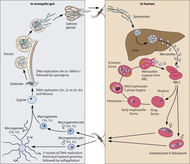
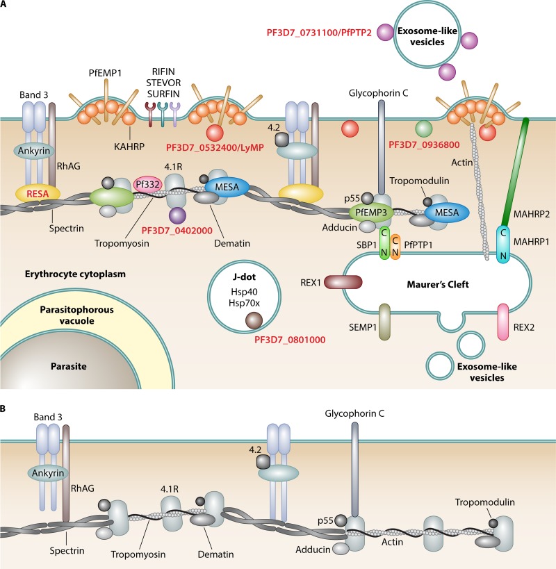
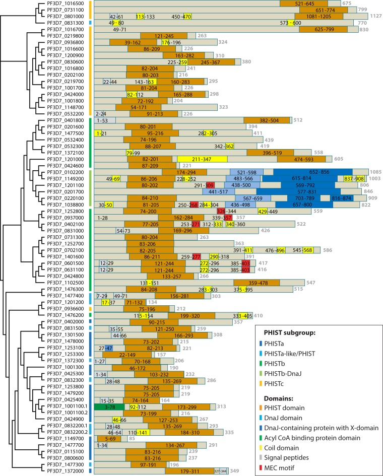
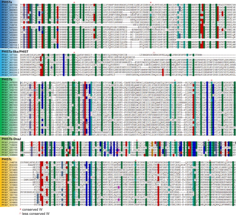
During the asexual cycle, Plasmodium falciparum extensively remodels the human erythrocyte to make it a suitable host cell. A large number of exported proteins facilitate this remodeling process, which causes erythrocytes to become more rigid, cytoadherent, and permeable for nutrients and metabolic products. Among the exported proteins, a family of 89 proteins, called the Plasmodium helical interspersed subtelomeric (PHIST) protein family, has been identified. While also found in other Plasmodium species, the PHIST family is greatly expanded in P. falciparum. Although a decade has passed since their first description, to date, most PHIST proteins remain uncharacterized and are of unknown function and localization within the host cell, and there are few data on their interactions with other host or parasite proteins. However, over the past few years, PHIST proteins have been mentioned in the literature at an increasing rate owing to their presence at various localizations within the infected erythrocyte. Expression of PHIST proteins has been implicated in molecular and cellular processes such as the surface display of PfEMP1, gametocytogenesis, changes in cell rigidity, and also cerebral and pregnancy-associated malaria. Thus, we conclude that PHIST proteins are central to host cell remodeling, but despite their obvious importance in pathology, PHIST proteins seem to be understudied. Here we review current knowledge, shed light on the definition of PHIST proteins, and discuss these proteins with respect to their localization and probable function. We take into consideration interaction studies, microarray analyses, or data from blood samples from naturally infected patients to combine all available information on this protein family.


Figures

FIG 1
FIG 1
Life cycle of P. falciparum. (Right) Upon the bite of a Plasmodium falciparum-infected female Anopheles mosquito, sporozoites are injected into the dermal tissue of the human host. Sporozoites quickly enter the bloodstream and are transported to the liver, where they invade liver cells and develop into liver schizonts. Through merosomes, thousands of merozoites are released into the bloodstream, where they invade erythrocytes, starting the asexual replication cycle. Each cycle, a few parasites cease replicating, commit to the sexual cycle, and develop into gametocytes. (Left) Mature gametocytes are taken up by a mosquito during a blood meal; rapidly develop into male and female gametes, which fuse together in the gut of the mosquito; form an ookinete that penetrates the gut wall; and undergo sexual replication in the oocyst, producing thousands of sporozoites. At the end, sporozoites migrate to the salivary gland and are ready to be transmitted to a human host during the next blood meal. (Adapted from reference .)
FIG 2
FIG 2
PHIST proteins in the remodeled iRBC. (A) Remodeled iRBC. During the asexual blood stage of P. falciparum, human erythrocytes are subject to extensive remodeling. All erythrocyte proteins are shown in gray (reviewed in reference 59). Parasite structures, compartments, and organelles are labeled in boldface type. PHIST proteins are labeled in red boldface type, and all parasite proteins are represented by colored shapes. References for PHIST proteins are indicated in the text. Knobs are parasite-derived protrusions in the host cell membrane, with knob-associated histidine-rich protein (KAHRP) being a prominent protein of these structures (149, 150). Maurer's clefts (reviewed in reference 151) are parasite-derived membranous structures in the iRBC cytoplasm involved in protein trafficking and are connected with knobs via actin filaments (152, 153). Maurer's cleft-associated histidine-rich protein 1 (MAHRP1) is a Maurer's cleft-resident protein (154) that potentially interacts with MAHRP2, the tether protein anchoring Maurer's clefts to the iRBC membrane (155). REX1 (156), REX2 (107), SEMP1 (157), SBP1 (158), and PfPTP1 (159) are other exported parasite proteins that localize to Maurer's clefts, with the latter two being located in a high-molecular-weight complex (159) and SBP1 interacting with the erythrocyte cytoskeleton proteins spectrin and band 4.1R (160). J dots are mobile, dot-like structures in iRBCs (161, 162). PfPTP2 is associated with exosomes, which are parasite-derived vesicles that are involved in cell-cell communication between iRBCs (43). RhAG, rhesus-associated antigen. (B) Cytoskeleton of an uninfected red blood cell (reviewed in reference 59). (Inspired by references and .)
FIG 3
FIG 3
Phylogenetic tree and protein domain prediction for PHIST proteins. The amino acid sequences of all 89 PHIST proteins were obtained from PlasmoDB. Of 19 PHIST proteins annotated as pseudogenes, 16 contained one or more premature stop codons in the amino acid sequence and were removed from the list. All those of the remaining 73 PHIST proteins with a PEXEL motif were PEXEL cleaved in silico by using the PEXEL motifs provided by Sargeant et al. (15), Boddey et al. (11), or Schulze et al. (12). The amino acid sequences were aligned with MUSCLE (163). The alignment is represented in a phylogenetic tree using the phylogenic tree tool built into MUSCLE at the EMBL-EBI website (164). The branch lengths are drawn in cladogram style and do not represent actual phylogenetic distances. The colored bars next to the gene identifications represent different PHIST subgroups, as indicated. The structure of the amino acid sequences was then analyzed with InterPro (https://www.ebi.ac.uk/interpro/) (165). A schematic representation of the results for each PHIST protein is shown next to its respective gene identification. The following different domains are highlighted: the PHIST domain, the DnaJ domain as defined by Pfam (family PF00226), the DnaJ-containing protein with an X domain as defined by InterPro (accession number IPR026894) or Pfam (family PF14308), the acyl coenzyme A (CoA) binding protein domain as defined by Pfam (family PF00887), coil domains as identified by InterPro, signal peptides as defined by SignalP or transmembrane domains, and the MEC motif as defined by Kilili and LaCount (39).
FIG 4
FIG 4
Sequence alignment of PHIST proteins. The processed amino acid sequences as described in the legend of Fig. 3 were sorted into PHIST subgroups, and individual alignments for each subgroup were obtained with CLUSTAL (166). The output was analyzed by using BioEdit (167). Conserved amino acids at a threshold of 83% are highlighted. Shown is the core of the alignment using the position of the first conserved tryptophan to align the different alignment blocks of the subgroups. Tryptophan residues conserved above or below the 83% threshold are marked as indicated.
FIG 5
FIG 5
PHIST domain modeling. (A) Visual representation of the positions of the conserved tryptophan residues of the PHIST subgroups, treating PHISTb and PHISTb-DnaJ as well as PHISTa and PHISTa-like/PHIST domains as separate subgroups to show the slight variations between them. A consensus sequence was generated for each subgroup alignment from Fig. 4. The consensus sequence was then split into blocks of 10 residues represented by gray boxes (boxes containing no tryptophans were shortened). The positions of the tryptophan residues conserved within a subgroup are marked by yellow bars. (B) To exemplify the differences in structure prediction, the Jpred4 (168, 169) output is shown for the PHIST protein PF3D7_0532400. The top sequence is the consensus sequence used for PHIST identification by Sargeant et al. (15). JNetPRED displays the consensus prediction: helices are marked with red tubes. JNetCONF provides confidence estimates for the prediction. High values mean high confidence. For the JNetHMM profile-based prediction, the six predicted helices are marked as red tubes. For the JNETPSSM-based prediction, the four helices are marked as red tubes, and sheets are marked with a green arrow.
FIG 6
FIG 6
Heat maps. (A) Heat map of phist gene expression of the P. falciparum 3D7 strain over the course of 53 h. Microarray data on transcript abundance were reported previously (46) (accessed through PlasmoDB, release 28, 31 March 2016). The heat map was constructed by using TMeV4.9 (170). phist genes were clustered into their respective subgroups and were then ordered by gene identification. Genes annotated “phista-like” or as “phist” genes were grouped together as “phist others.” Yellow boxes to the right of the heat map indicate PHIST proteins present in the early gametocyte proteome (35), differentially expressed phist genes in pregnancy-associated malaria (29, 49, 50, 84, 92, 112) and in cerebral malaria (32, 34), or if variant expression has been reported (55). Green to red colors represent fold changes (log fold changes from −3 to 3). (B) For a number of phist genes, no expression data were available in the data set, and these genes were grouped first by PHIST subgroup and then by gene identification. Additional information for selected PHIST proteins is provided in Tables 1 to 5. The colored bars next to the gene identifications indicate the PHIST subgroup.
None
None
None

Similar articles

Cited by

References

    1. Boddey JA, Cowman AF. 2013. Plasmodium nesting: remaking the erythrocyte from the inside out. Annu Rev Microbiol 67:243–269. doi:10.1146/annurev-micro-092412-155730. - DOI - PubMed
    1. Tuteja R. 2007. Malaria—an overview. FEBS J 274:4670–4679. doi:10.1111/j.1742-4658.2007.05997.x. - DOI - PubMed
    1. WHO. 2015. World malaria report 2015. WHO, Geneva, Switzerland.
    1. Amino R, Giovannini D, Thiberge S, Gueirard P, Boisson B, Dubremetz JF, Prevost MC, Ishino T, Yuda M, Menard R. 2008. Host cell traversal is important for progression of the malaria parasite through the dermis to the liver. Cell Host Microbe 3:88–96. doi:10.1016/j.chom.2007.12.007. - DOI - PubMed
    1. Sturm A, Amino R, van de Sand C, Regen T, Retzlaff S, Rennenberg A, Krueger A, Pollok JM, Menard R, Heussler VT. 2006. Manipulation of host hepatocytes by the malaria parasite for delivery into liver sinusoids. Science 313:1287–1290. doi:10.1126/science.1129720. - DOI - PubMed

Publication types

MeSH terms

Substances

LinkOut - more resources