Review
. 2016 Dec;16(24):3111-3125.
doi: 10.1002/pmic.201600116.

Key regulators of galectin-glycan interactions

Affiliations
Review

Key regulators of galectin-glycan interactions

Nourine A Kamili et al. Proteomics. 2016 Dec.

Abstract

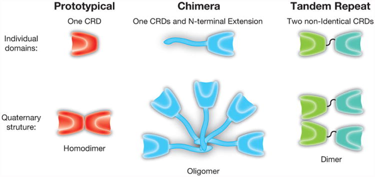
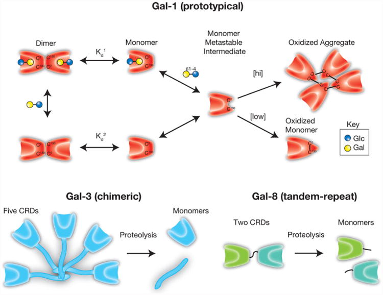
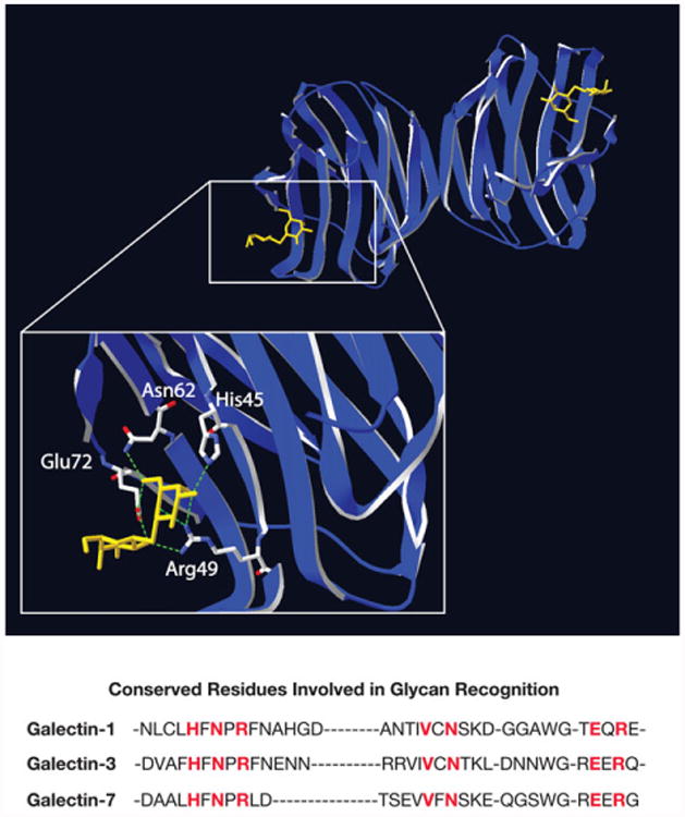
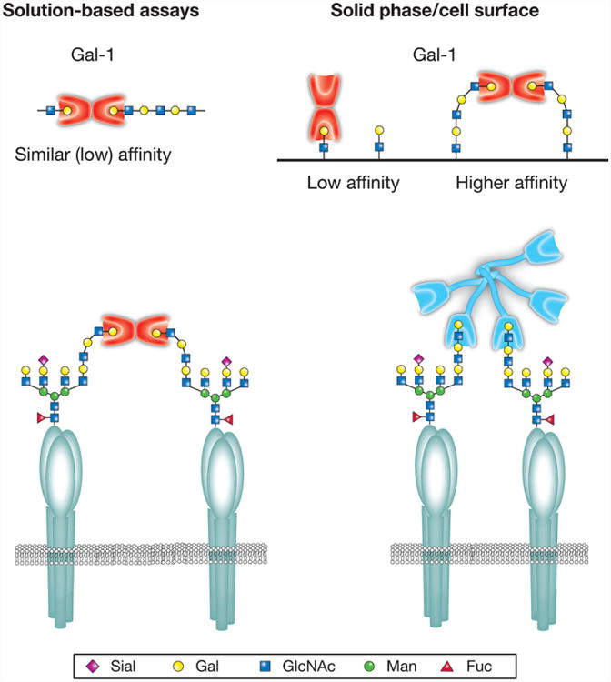
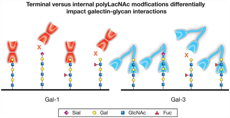
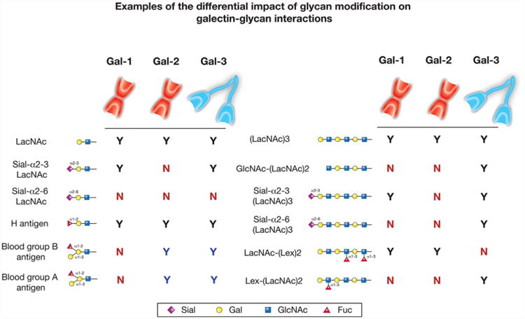
Protein-ligand interactions serve as fundamental regulators of numerous biological processes. Among protein-ligand pairs, glycan binding proteins (GBPs) and the glycans they recognize represent unique and highly complex interactions implicated in a broad range of regulatory activities. With few exceptions, cell surface receptors and secreted proteins are heavily glycosylated. As these glycans often represent highly regulatable post-translational modifications, alterations in glycosylation can fundamentally impact GBP recognition. Among GBPs, galectins in particular appear to engage a diverse set of glycan determinants to impact a broad range of biological processes. In this review, we will explore factors that impact galectin activity, including the effect of glycan modification on galectin-glycan interactions.

Keywords: Frontal Affinity Chromatography; Galectin; Glycan; Glycan Binding Protein; Glycoproteomics; Surface Plasmon Resonance.


Figures

Figure 1
Figure 1
Galectin family members. Galectins can be divided into three subfamilies, based on the organization of the carbohydrate recognition domain: prototypical (galectin-1, galectin-2, galectin-7 and galectin-10), chimeric (galectin-3) and tandem repeat (galectin-4, galectin-8, galectin-9 and galectin 12). Each of these subfamilies also exhibit unique quaternary structures, with prototypical and at least some tandem repeat galectins forming dimers and chimeric galectin-3 forming higher order oligomers.
Figure 2
Figure 2
Regulation of galectin activity. Galectin-1 (Gal-1) can undergo oxidative inactivation through the formation of intramolecular or intermolecular disulfide bond formation, depending on Gal-1 concentration. In contrast, galectin-3 (Gal-3), which is resistant to oxida-tive inactivation, can be inactivated by proteolytic cleavage of the N-terminal domain required for Gal-3 oligomerization. Similar cleavage of the intervening linker between the carbohydrate recognition domains of tandem-repeat galectins, as shown for galectin-8 (Gal-8), can also eliminate their biological activity.
Figure 3
Figure 3
Structure of galectin-1 carbohydrate recognition domain. Prototypical galectins exist as homodimers with carbohydrate recognition domains that face in opposite directions. Structural studies elucidated the carbohydrate binding pocket of the galectin family, highlighting conserved amino acids responsible for the β-galactoside specificity of galectins. Lactosamine modification can differential impact core carbohydrate recognition domain interactions with β-galactoside ligands. Here, galectin-1 is shown associated with its inhibitor TDG. The crystal structure data was obtained from open source NCI database (PDBID 3OYW) and reconstructed using SwissPBD Viewer. Key residues involved in glycan recognition for galectin-1, galectin-3 and galectin-7 are highlighted in red.
Figure 4
Figure 4
Quaternary structure can influence galectin–glycan interactions. While the majority of human galectins exhibit a preference for polylactosamine (polyLacNAc) glycans in solid-phase assays, this predilection is not observed for some galectins in solution-based approaches. This may in part reflect different modes of polyLacNAc recognition coupled with unique aspects of the quaternary structure of individual galectin family members. Galectin-1 (Gal-1) and other prototypical galectins primarily exist as rigid dimers with carbohydrate recognition domains facing opposite directions. Several prototypical galectins also appear to preferentially engage the terminal lactosamine of polyLacNAc. As a result, co-engagement of lactosamine-containing ligands is likely facilitated by the conformational flexibility afforded by extended polyLacNAc structures. In contrast, in solution-based assays, crosslinking interactions appreciated on solid supports are no longer apparent. Unlike Gal-1, galectin-3 (Gal-3) and other tandem repeat galectins oligomerize through flexible linkers and therefore do not exhibit the same rigid carbohydrate recognition domain orientation. These galectins appear to have instead evolved the ability to primarily interact with polyLacNAc through internal lactosamine motifs, allowing the same polyLacNAc preference to be observed in solution and solid-phase-based assays. These differences in polyLacNAc engagement allow simple polyLacNAc modifications to differentially impact galectin–glycan interactions with significant consequences on cellular sensitivity to galectin-induced signaling.
Figure 5
Figure 5
Individual galectin family members differentially recognize polylactosamine (polyLacNAc). While each galectin family member displays a common ability to interact with polyLacNAc, the mode of interaction between different family members can fundamentally differ. For example, galectin-1 (Gal-1) preferentially engages the terminal lactosamine (LacNAc) unit of polyLacNAc glycans. Consequently, terminal modifications, such as α2,6-sialylation, are more likely to impact recognition by Gal-1. In contrast, galectin-3 (Gal-3) preferentially engages internal LacNAc units within polyLacNAc and therefore may not be impacted by terminal modifications, but may instead be influenced by internal polyLacNAc modifications.
Figure 6
Figure 6
Lactosamine modification differentially impacts galectin binding. While galectin proteins are defined by their common affinity for β-galactoside, each galectin varies in its recognition of modified galactose residues. For galectin-1 (Gal-1), galectin-2 (Gal-2) and galectin-3 (Gal-3), the affinity for modified lactosamine (LacNAc) motifs compared to LacNAc alone is shown on the left panel. Similarly, binding preferences toward different polylactosamine (polyLacNAc) modifications is compared to polyLacNAc in the right panel. The black Ys indicate positive binding between each respective galectin and the glycan structure, while the blue Ys indicate increased binding compared to baseline binding (LacNAc for the left panel and polyLacNAc for the right panel). Gal-1, Gal-2 and Gal-3 all display higher binding toward polyLacNAc than LacNAc in solid-phase assays. The red Ns indicate no binding toward the respective glycan.

References

    1. Marth JD, Grewal PK. Mammalian glycosylation in immunity. Nat Rev Immunol. 2008;8:874–887. - PMC - PubMed
    1. Cummings RD. The repertoire of glycan determinants in the human glycome. Mol Biosyst. 2009;5:1087–1104. - PubMed
    1. Rabinovich GA, van Kooyk Y, Cobb BA. Glycobiology of immune responses. Ann NY Acad Sci. 2012;1253:1–15. - PMC - PubMed
    1. Rabinovich GA, Toscano MA. Turning ‘sweet’ on immunity: galectin-glycan interactions in immune tolerance and inflammation. Nat Rev Immunol. 2009;9:338–352. - PubMed
    1. Liu FT, Rabinovich GA. Galectins as modulators of tumour progression. Nat Rev Cancer. 2005;5:29–41. - PubMed

LinkOut - more resources