. 2016 Sep 30;119(8):e110-26.
doi: 10.1161/CIRCRESAHA.116.308929. Epub 2016 Aug 31.

Platelet and Erythrocyte Sources of S1P Are Redundant for Vascular Development and Homeostasis, but Both Rendered Essential After Plasma S1P Depletion in Anaphylactic Shock

Affiliations

Platelet and Erythrocyte Sources of S1P Are Redundant for Vascular Development and Homeostasis, but Both Rendered Essential After Plasma S1P Depletion in Anaphylactic Shock

Salomé L Gazit et al. Circ Res. .

Abstract

Rationale: Sphingosine-1-phosphate (S1P) signaling is essential for vascular development and postnatal vascular homeostasis. The relative importance of S1P sources sustaining these processes remains unclear.

Objective: To address the level of redundancy in bioactive S1P provision to the developing and mature vasculature.

Methods and results: S1P production was selectively impaired in mouse platelets, erythrocytes, endothelium, or smooth muscle cells by targeted deletion of genes encoding sphingosine kinases -1 and -2. S1P deficiency impaired aggregation and spreading of washed platelets and profoundly reduced their capacity to promote endothelial barrier function ex vivo. However, and in contrast to recent reports, neither platelets nor any other source of S1P was essential for vascular development, vascular integrity, or hemostasis/thrombosis. Yet rapid and profound depletion of plasma S1P during systemic anaphylaxis rendered both platelet- and erythrocyte-derived S1P essential for survival, with a contribution from blood endothelium observed only in the absence of circulating sources. Recovery was sensitive to aspirin in mice with but not without platelet S1P, suggesting that platelet activation and stimulus-response coupling is needed. S1P deficiency aggravated vasoplegia in this model, arguing a vital role for S1P in maintaining vascular resistance during recovery from circulatory shock. Accordingly, the S1P2 receptor mediated most of the survival benefit of S1P, whereas the endothelial S1P1 receptor was dispensable for survival despite its importance for maintaining vascular integrity.

Conclusions: Although source redundancy normally secures essential S1P signaling in developing and mature blood vessels, profound depletion of plasma S1P renders both erythrocyte and platelet S1P pools necessary for recovery and high basal plasma S1P levels protective during anaphylactic shock.

Keywords: anaphylaxis; endothelium; shock; sphingosine-1-phosphate; vascular endothelial function; vascular permeability; vascular tone regulation.


Figures

Figure 1
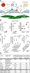
Figure 1. Generation of S1P-deficient mice
A. Schematic overview of S1P signaling in the vasculature. S1P receptors on endothelial cells and possibly smooth muscle cells play important roles in regulating vascular development, endothelial barrier function and vascular tone. Multiple cell types provide S1P to plasma, where it binds to HDL, albumin, and LDL. B-C. S1P concentrations as determined by HPLC and LC-MS/MS in lysed platelets, supernatants of washed and activated platelets, erythrocytes, plasma and/or serum from mice with single global deficiency or compound tissue-specific deficiency in Sphk1&2 in megakaryocytes (Sphk1f/-:2f/-:Pf4Cre+), blood endothelium (Sphk1f/-:2f/-:PdgfbiCre+), hematopoietic cells, hepatocytes and lymphatic endothelium (Sphk1f/-:2f/-:Mx1Cre+) or the latter combined (Sphk1f/-:2f/-:Mx1Cre+:PdgfbiCre+). Compound gene deletion was performed either by tissue-specific recombination of one loxP-flanked copy of each kinase gene in a context where the other copy was deleted in all cells (f/-:f/-) or by tissue-specific recombination of two loxP flanked copies of one kinase gene in a context where both copies of the other kinase gene were deleted in all cells (f/f:-/-). D. Overview of Cre alleles, their target cells, and effect of Sphk deletion on S1P levels in platelets, erythrocytes and plasma under homeostasis as determined above or published elsewhere., , ND, not determined. Note that erythrocytes and platelets make near exclusive use of Sphk1 and Sphk2, respectively, for S1P production, and that while relevant target cells of Mx1-Cre (erythrocytes and lymphatic endothelial cells) continuously provide S1P to plasma, platelets only release their S1P content upon activation and thus contribute to serum but not plasma S1P. Blood endothelium does not contribute measurably to plasma S1P, but mays still constitute a relevant source for activation of vascular receptors.
Figure 2
Figure 2. Platelet S1P deficiency impairs functional responses in washed platelets
The function of Sphk 1&2 deficient platelets was addressed ex vivo using washed platelets by assessing (A) α-thrombin (10 nM)- induced changes in cytosolic [Ca2+] by fura-2 ratiometric analysis (B) PAR4-AP (200 μM) - induced P-selectin exposure by flow cytometry, (C) collagen (0.75 μg/mL) and ADP (1.5 μM) - induced aggregation by densitometry (AUC, area under the curve), and (D) spreading on fibrinogen. Sphk 1&2-deficient platelets show normal calcium signaling, slightly delayed P-Selectin exposure, delayed aggregation to collagen (difference observed at 1 but not 3 minutes) and reduced aggregation to ADP and spreading on fibrinogen (red) relative to platelets from littermate control mice (black). Platelet adhesion to fibrinogen and spreading on BSA were not different between the groups.
Figure 3
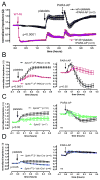
Figure 3. S1P is critical for the capacity of platelets to promote endothelial barrier function ex vivo
HUVEC were cultured on gold electrodes and electrical impedance, reflecting transendothelial electrical resistance and barrier function, measured. After an initial stabilization period, 10 washed platelets from wild-type (A) or indicated Sphk deficient (B-D) mice or littermate controls were added to HUVEC monolayers (B-D, left panel), and then activated with PAR4-AP (500 μM) (B-D, right panel). Endothelial cells in (A) were treated with the S1P1 antagonist W146 (1 μM, purple) or vehicle control (black) for 1 hour prior to the addition of platelets. In A, impedance was normalized before addition of W146 or vehicle control. In B-E, impedance was normalized before platelet addition in left panels and before PAR4 addition in right panels. The PAR4-AP did not significantly alter barrier function in HUVECs in the absence of platelets, and platelets alone had negligible effects on impedance of fibronectin- or collagen- coated electrodes.
Figure 4
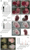
Figure 4. Platelet- and erythrocyte-derived S1P are dispensable for developmental blood-lymph separation and vascular integrity
A. Embryos with defective thrombopoiesis (Nf-e2-/-) or rendered deficient in megakaryocyte Sphk1 in a Sphk2-/- background (Sphk1f/f:2-/-:Pf4Cre+) were recovered at E14.5 and inspected for the presence of blood in lymphatic vessels (white arrow) and intracranial hemorrhage (hatched white arrow). Note the presence of both phenotypes in Nf-e2-/- embryos but not littermate controls or platelet S1P deficient embryos. While embryos rendered deficient in erythrocyte Sphk1 in a Sphk2-/- background (Sphk1f/f:2-/-:EporCre+) showed widespread bleeding (black arrowhead) and in some cases exencephaly (asterisk) at E12.5, this was not observed when Sphk1 was deleted in erythrocytes in a Sphk2+/- background (Sphk1f/f:2f/-:EporCre), nor when Sphk1&2 were both deleted specifically in erythrocytes (Sphk1f/-:2f/-:EpoRCre+), or when both erythrocyte and platelet sources were impaired (Sphk1-/-:2f/(f)-:Pf4Cre+). The left panel summarizes observations in collected embryos. B. Sphk deficient and Nf-e2-/- neonates were sacrificed on postnatal day 0-2 and brains dissected after transcardial PBS perfusion to look for evidence of hemorrhage. Left panel: representative brains. Right panel: summary. No significant difference in bleeding prevalence was observed with platelet- or platelet- and erythrocyte-S1P deficiency, whereas bleeding was significantly more frequent in Nf-e2-/- than in corresponding littermate controls. Images were acquired in PBS immersion with a Leica M165FC stereo microscope.
Figure 5
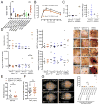
Figure 5. Effects of S1P deficiencies on vascular integrity, hemostasis, thrombosis, and leukocyte recruitment in adult mice
A. Constitutive Evans Blue leak to lung in mice deficient in S1P sources or the endothelial S1P1 receptor. B. Histamine (60 mg) –induced paw swelling normalized for increase in paw diameter after vehicle injection in the contralateral paw. C. Hemoglobin and myeloid cells in bronchoalveolar lavage 6 hours after intranasal inoculation of LPS (25 mg). Platelet depletion triggered bleeding and reduced leukocyte accumulation in lung, whereas S1P deficiency did not affect either parameter. D. Hemoglobin, bleeding score, myeloperoxidase (MPO) activity and weight of skin biopsies 4 hours after semi-simultaneous s.c. injection of anti-BSA and i.v. injection of BSA (left panel). Bleeding, MPO activity, and weight were all increased in rpA biopsies relative to control independent of genotype. Representative images (right panel) show occasional mild petechiae at sites injected with anti-BSA antibody in Sphk1f/-:2f/- mice independent of Pf4-Cre status. E-F Bleeding times after removal of the distal 3 mm of the tail (E) and time to arterial occlusion after 4 minutes exposure of the carotid artery to a FeCl3 (15%) swab (F). Platelet S1P deficiency did not influence either endpoint, nor hemoglobin content in collected blood in E or in embolization in F. G. C57BL/6 wild-type mice were transplanted with platelet S1P deficient bone marrow (BM) or Nf-e2-/- fetal liver (FL) cells the day after Cesium 137 irradiation. 10 or 13 days (d) later, brains were removed after transcardial perfusion. Left panel, representative brains 13 days after transplantation; right panel, summary. Platelet- but not platelet S1P-deficiency resulted in brain bleeding after irradiation.
Figure 6
Figure 6. Platelets and S1P, but not platelet-derived S1P, are essential for lymph node hemostasis after immunization
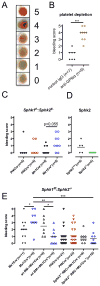
A-E. Mice were immunized in the right hock and draining and control LNs isolated one week later for gross assessment of bleeding. Bleeding on the efferent surface of the right popliteal lymph node was scored in a blinded manner according to the scale shown in A. In B, anti-GPIbα antibodies were administered to C57BL/6 males the day before harvest, and thrombocytopenia confirmed immediately before transcardial perfusion. Thrombocytopenia induced bleeding in draining lymph nodes (B) while deficiency in platelet-derived S1P (Sphk1f/-:2f/-:Pf4Cre+ or Sphk2-/-) did not. C-D. Lack of all circulating sources of S1P (Sphk1f/-:2f/-:Mx1Cre+) was associated with a bleeding tendency (C). In a background globally deficient in Sphk2 (E), deletion of Sphk1 in hematopoietic cells and lymphatic endothelial cells (Sphk1f/f:2-/-:Mx1Cre+), but not in megakaryocytes (Sphk1f/f:2-/-:Pf4Cre+), was associated with lymph node bleeding. Sensitization to bleeding associated with plasma S1P deficiency could be rescued by transplantation of wild-type bone marrow (wt BM) cells or by transfusions of erythrocytes from Sphk2-/- donors (E). Images were acquired in PBS immersion with a Leica M165FC stereo microscope.
Figure 7
Figure 7. Plasma S1P depletion renders otherwise redundant S1P sources essential for recovery from anaphylactic shock
A. Intravital imaging of interactions between platelets, leukocytes, and the vessel wall before and 20 minutes after PAF (4 μg/kg)-induced anaphylaxis. Images extracted from time-lapse microscopy videos in the Online Supplement. PAF injection led to transient interactions between platelets, leukocytes and the vessel wall in both arteries and veins, as well as to firm adhesion (≥ 5 sec). White and black arrows indicate firmly adherent platelets and leukocytes, respectively. Insets show higher-magnification views of squared regions. Images are representative of observations made in 6 mice. B. S1P concentrations in platelets, erythrocytes and plasma of C57BL/6J wild-type mice 40 minutes after PAF (4 μg/kg) or vehicle challenge. Note S1P depletion from platelets and plasma, but not erythrocytes. C-F. Mice were injected with PAF at indicated concentrations and survival monitored for 60 minutes. Note that S1P deficiency in circulating cells; in chimeric wild-type (wt) mice with Sphk deficiency in all bone marrow (BM) cells (Sphk1f/-:2f/-:Mx1Cre+), or selectively in megakaryocytes (Sphk1f/-:2f/-:Pf4Cre+) or mice lacking S1P production in erythrocytes (Sphk1f/f:+/-:EporCre+) all showed a significant increase in mortality relative to controls (C). By contrast, S1P deficiency in the vessel wall, either in blood endothelium (Sphk1f/-:2f/-:PdgfbiCre+) or in smooth muscle cells (Sphk1f/-:2f/-:Sm22Cre+) did not increase mortality in isolation, although deficiency in blood endothelium significantly increased mortality in the context of pan-hematopoietic deficiency (Sphk1f/-:2f/-:PdgfbiCre+:Mx1Cre+) (D). Aspirin significantly increased mortality in wild-type mice, but not in chimeric wild-type mice with Sphk1f/f:2-/-Pf4Cre+ BM (E). A higher PAF dose was used in the latter because of protective effects of Sphk2 deficiency (F), which may be conferred by high plasma S1P levels in these mice.
Figure 8
Figure 8. S1P promotes recovery from anaphylactic shock by S1P2-mediated enhancement of vascular tone
A. Mice selectively lacking S1P1 in blood endothelium (S1pr1f/f:PdgfbiCre+) were challenged with PAF and survival monitored for 60 minutes. B. Plasma S1Pless (Sphk1f/-:2f/-:Mx1Cre+), S1P2 deficient mice (S1pr2-/-) and littermate controls were challenged with PAF (2 μg/kg) under pentobarbital anesthesia and blood pressure and heart rate monitored for 60 minutes and normalized to pre-stimulation values. Asterisk indicates death of an animal. C. Plasma S1Pless (Sphk1f/f:2-/-:Mx1Cre+), chimeric wild-type (wt) mice with megakaryocyte Sphk1&2 deficient bone marrow (BM)(Sphk1f/-:2f/-:Pf4Cre+), S1P2 deficient mice (S1pr2-/-) and respective controls were challenged with PAF (4 μg/kg) and left ventricular systolic function and blood flow velocities in the right renal artery and basilar trunk determined 30 minutes later by echocardiography and ultrasound pulsed Doppler analysis under light (0.5 %) isoflurane anesthesia. Upper panel: left ventricular internal diameter in diastole (LVIDd), cardiac output, right renal artery and basilar trunk mean blood flow velocities before and after PAF challenge. D-F. Mice globally deficient in S1P2 either alone (S1pr2-/-; D) or in conjunction with Sphk2 (S1pr2-/:Sphk2-/-; E) or S1P3 (S1pr2-/:S1pr3-/-; F) and littermate controls were challenged with PAF at indicated concentrations and survival monitored for 60 minutes. Note that only S1P2 deficiency sensitizes to PAF, independent of S1P3, although residual protection by Sphk2 deficiency in a S1pr2-/- background suggests the involvement of second receptor.

References

    1. Proia RL, Hla T. Emerging biology of sphingosine-1-phosphate: Its role in pathogenesis and therapy. J Clin Invest. 2015;125:1379–1387. - PMC - PubMed
    1. Spiegel S, Milstien S. Sphingosine-1-phosphate: An enigmatic signalling lipid. Nat Rev Mol Cell Biol. 2003;4:397–407. - PubMed
    1. Kihara Y, Maceyka M, Spiegel S, Chun J. Lysophospholipid receptor nomenclature review: Iuphar review 8. Br J Pharmacol. 2014;171:3575–3594. - PMC - PubMed
    1. Liu Y, Wada R, Yamashita T, Mi Y, Deng CX, Hobson JP, Rosenfeldt HM, Nava VE, Chae SS, Lee MJ, Liu CH, Hla T, Spiegel S, Proia RL. Edg-1, the g protein-coupled receptor for sphingosine-1-phosphate, is essential for vascular maturation. J Clin Invest. 2000;106:951–961. - PMC - PubMed
    1. Jung B, Obinata H, Galvani S, Mendelson K, Ding BS, Skoura A, Kinzel B, Brinkmann V, Rafii S, Evans T, Hla T. Flow-regulated endothelial s1p receptor-1 signaling sustains vascular development. Dev Cell. 2012;23:600–610. - PMC - PubMed

MeSH terms