Review
. 2016 Sep 1:2:16059.
doi: 10.1038/nrdp.2016.59.

Infective endocarditis

Affiliations
Review

Infective endocarditis

Thomas L Holland et al. Nat Rev Dis Primers. .

Abstract

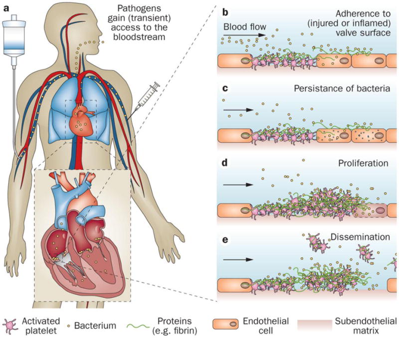
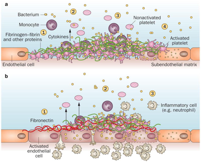
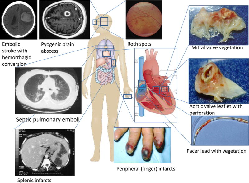
Infective endocarditis (IE) is a rare, life-threatening disease that has long-lasting effects even among patients who survive and are cured. IE disproportionately affects those with underlying structural heart disease and is increasingly associated with health care contact, particularly in patients who have intravascular prosthetic material. In the setting of bacteraemia with a pathogenic organism, an infected vegetation may form as the end result of complex interactions between invading microorganisms and the host immune system. Once established, IE can involve almost any organ system in the body. The diagnosis of IE may be difficult to establish and a strategy that combines clinical, microbiological and echocardiography results has been codified in the modified Duke criteria. In cases of blood culture-negative IE, the diagnosis may be especially challenging, and novel microbiological and imaging techniques have been developed to establish its presence. Once diagnosed, IE is best managed by a multidisciplinary team with expertise in infectious diseases, cardiology and cardiac surgery. Antibiotic prophylaxis for the prevention of IE remains controversial. Efforts to develop a vaccine that targets common bacterial causes of IE are ongoing, but have not yet yielded a commercially available product.


Conflict of interest statement

V.G.F. reports the following potential conflicts of interest: Chair of the Scientific Advisory Board for Merck V710 Staphylococcus aureus vaccine; paid consultant for Pfizer, Novartis, Galderma, Novadigm, Durata, Debiopharm, Genentech, Achaogen, Affinium, Medicines Co., Cerexa, Tetraphase, Trius, MedImmune, Bayer, Theravance, Cubist, Basilea and Affinergy; grants pending from MedImmune, Actavis/Forest/Cerexa, Pfizer, Merck/Cubist, Advanced Liquid Logics, Theravance and Novartis; royalties from UpToDate; personal fees for development or presentation of educational presentations from Green Cross, Cubist, Cerexa, Durata and Theravance,; and a patent pending related to sepsis diagnostics. T.L.H. reports the following potential conflicts of interest: paid consultant for The Medicines Company and Basilea Pharmaceutica; and royalties from UpToDate. A.S.B. reports the following potential conflicts of interest: Research Grants from ContraFect and Theravance; and Advisory Board member for ContraFect. J.M.M. reports the following potential conflicts of interest: consulting honoraria and/or research grants from AbbVie, Bristol-Myers Squibb, Cubist, Genentech, Merck, Novartis, Gilead Sciences and ViiV Healthcare. The other authors declare no potential conflicts of interest.
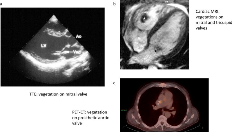
Figures

Figure 1
Figure 1. Global epidemiology of causative pathogens involved in endocarditis
The causative agents of infective endocarditis differ geographically. Data derived from Murdoch et al. CoNS, Coagulase-negative staphylococci; HACEK, Haemophilus species, Aggregatibacter species, Cardiobacterium hominis, Eikenella corrodens, and Kingella species; Strep, streptococcal species; VGS, viridans group streptococci.
Figure 2
Figure 2. Pathogenesis of endocarditis
a. Pathogens gain access to the bloodstream, for example via an intravenous catheter, injection drug use or from a dental source. b Pathogens adhere to an area of abnormal cardiac valve surface. c Some pathogens, such as S. aureus, obtain intracellular access to the valve endothelium. d The infected vegetation is created by burying of the proliferating organism within a protective matrix of serum molecules. e –Vegetation particles can detach and disseminate to form emboli. These may lead to complications such as ischemic stroke, mycotic aneurysms and infarcts or abscesses at remote sites. Figure adapted from Werdan et al. Mechanisms of infective endocarditis: pathogen-host interaction and risk states. Nat Rev Cardiol. 2014;11:35–50.
Figure 3
Figure 3. Mechanisms of infective endocarditis
a Valve colonization as a consequence of mechanical injury. 1) Nonbacterial thrombotic endocarditis. 2) Bacteria bind to coagulum and colonize it during transient bacteraemia. Adhered monocytes release tissue factor and cytokines. 3) More platelets are attracted and become activated and the vegetation grows. 4) Endothelial cells are infected and can be lysed by bacterial products, or bacteria can persist inside the cells. b Valve colonization as a consequence of an inflammatory endothelial lesion. 1) Activated endothelial cells express integrins that promote the local deposition of fibronectin; bacteria such as S. aureus adhere to this protein. 2) Bacteria are internalized and endothelial cells release tissue factor and cytokines, causing blood clotting and promoting the extension of inflammation and vegetation formation. 3) Infected endothelial cells can be lysed by bacterial products or bacteria can persist inside the cells. Figure adapted from Werdan et al. Mechanisms of infective endocarditis: pathogen-host interaction and risk states. Nat Rev Cardiol. 2014;11:35–50.
Figure 4
Figure 4. End-organ manifestations of endocarditis
A) CT scans of pyogenic brain abscess and embolic stroke with haemorrhagic conversion. B) CT scan demonstrating multiple septic pulmonary emboli. C) CT scan demonstrating peripheral wedge-shaped splenic infarcts. D) Roth spots on funduscopic exam. E) Infarcts affecting multiple fingers. F) Explanted mitral valve with vegetation. G) Explanted aortic valve leaflet with vegetation and perforation. H) Pacemaker lead with vegetation. Roth spots photo courtesy of Walter B. Holland, MD.
Figure 5
Figure 5. Imaging modalities for diagnosis of endocarditis
a Transthoracic echocardiography demonstrating native mitral valve vegetation. b Cardiac MRI, systolic frame demonstrating vegetations in the sub-tricuspid valve chordal apparatus with adherent thrombus (white asterisk) and posterior mitral valve leaflet (black asterisk). c PET-CT – In this patient, infection of a prosthetic aortic valve was suspected but echocardiography was inconclusive. Using PET-CT, inflammatory leukocytes are visualized after taking up radiolabeled glucose, demonstrating an area of active infection on the aortic valve. Ao, aorta; LV, left ventricle; Veg, vegetation. Pacemaker lead images courtesy of Gail Peterson, MD.

References

    1. Contrepois A. Towards a history of infective endocarditis. Medical history. 1996;40:25–54. - PMC - PubMed
    1. Osler W. The Gulstonian Lectures, on Malignant Endocarditis. British medical journal. 1885;1:577–579. - PMC - PubMed
    1. Bin Abdulhak AA, et al. Global and regional burden of infective endocarditis, 1990–2010: a systematic review of the literature. Glob Heart. 2014;9:131–143. - PubMed
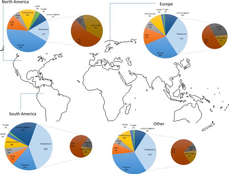
    1. Murdoch DR, et al. Clinical presentation, etiology, and outcome of infective endocarditis in the 21st century: the International Collaboration on Endocarditis-Prospective Cohort Study. Archives of internal medicine. 2009;169:463–473. This prospective cohort study of 2781 adults with definite endocarditis demonstrated that IE had shifted from a subacute disease of younger people with rheumatic valvular abnormalities, to one in which the presentation is more acute and is characterized by a high rate of S. aureus infection in patients with previous health care exposure. - PMC - PubMed
    1. Thayer W. Studies on bacterial (infective) endocarditis. Johns Hopkins Hosp Rep. 1926;22:1.