. 2016:2:16016.
doi: 10.1038/npjbcancer.2016.16. Epub 2016 Jun 15.

A microchip platform for structural oncology applications

Affiliations

A microchip platform for structural oncology applications

Carly E Winton et al. NPJ Breast Cancer. 2016.

Abstract

Recent advances in the development of functional materials offer new tools to dissect human health and disease mechanisms. The use of tunable surfaces is especially appealing as substrates can be tailored to fit applications involving specific cell types or tissues. Here we use tunable materials to facilitate the three-dimensional (3D) analysis of BRCA1 gene regulatory complexes derived from human cancer cells. We employed a recently developed microchip platform to isolate BRCA1 protein assemblies natively formed in breast cancer cells with and without BRCA1 mutations. The captured assemblies proved amenable to cryo-electron microscopy (EM) imaging and downstream computational analysis. Resulting 3D structures reveal the manner in which wild-type BRCA1 engages the RNA polymerase II (RNAP II) core complex that contained K63-linked ubiquitin moieties-a putative signal for DNA repair. Importantly, we also determined that molecular assemblies harboring the BRCA15382insC mutation exhibited altered protein interactions and ubiquitination patterns compared to wild-type complexes. Overall, our analyses proved optimal for developing new structural oncology applications involving patient-derived cancer cells, while expanding our knowledge of BRCA1's role in gene regulatory events.


Conflict of interest statement

The authors declare no conflict of interest.

Figures

Figure 1
Figure 1
The tunable microchip system captures native proteins produced in breast cancer cells. Native BRCA1 protein assemblies formed in the nucleus of hereditary breast cancer cells were tethered to tunable SiN-based microchips for 3D structural analysis. Representative 3D reconstructions (white and cyan) show variations in structural features and molecular domains as described in the present work.
Figure 2
Figure 2
EM structure reveals BRCA1 domains directly engage the RNAP II core proximal to DNA in human breast cancer cells. The EM density map (white) is shown in different orientations. The position of the BRCT domain was recently determined based on antibody-labeling results. In the present study, the BRCA1–BARD1 RING domain was uniquely placed into the density map. The DNA strand (blue) was positioned over the DNA channel accordingly. K63-linked ubiquitins occupied the remaining density. The RNAP II core was localized in the EM map based on a model of the RNAP II X-ray crystal structure. Bar = 5 nm. Cross-sections through the density map (1–4) indicate the overall fit of the atomic models within the envelope. Also see Supplementary Figure S3 and Supplementary Movie S1. BRCT, BRCA1 C-terminal domain; BARD1, BRCA1-associated ring domain protein; EM, electron microscopy; RNAP II, RNA polymerase II.
Figure 3
Figure 3
BRCA1 engages DNA in a variable manner while bound to the RNAP II core. (a) The composite EM structure was compared to transient intermediate structures having low and high DNA occupancies. (b) An intermediate structure having low DNA occupancy (yellow density map) accommodates a short fragment of DNA (blue) located proximal (black arrows) to the BRCA1–BARD1 RING domains. Limited density was present in the density map to accommodate K63-linked ubiquitins. (c) An intermediate structure having high DNA occupancy (cyan density map) accommodates a longer strand of DNA (blue) located proximal (black arrows) to the BRCA1–BARD1 RING domains. Bar=5 nm. Also see Supplementary Movie S2 and S3. BARD1, BRCA1-associated ring domain protein; EM, electron microscopy.
Figure 4
Figure 4
The pSer5 peptide exhibits the optimal stereochemistry to interact with the BRCT domain. (a) The composite 3D structure highlighting the BRCT domain (gray) within the density map. (b) A close-up view of the BRCT crystal structure (pdbcode, 3K0H ) showing that the hydrophobic binding pocket (gray rectangle) accommodates a known peptide, pSPTF. Molecular modeling experiments were performed to overlay the pSer5 and pSer2 peptides onto the pSPTF model. (c) The pSer5 peptide contains a terminal tyrosine residue (blue dashed circle) that fits within the hydrophobic binding cleft. Also see Supplementary Movie S4. (d) The pSer2 peptide contains a terminal serine residue (blue dashed circle) that does not maintain the proper stereochemistry to optimally fit within the BRCT binding site. BRCT, BRCA1 C-terminal domain.
Figure 5
Figure 5
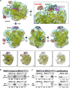
EM structure of mutated BRCA15382insC transcriptional complexes. (a) The EM density map (cyan) shown in different orientations was calculated using the RELION software package. Placement of the BRCA1–BARD1 (magenta, green) RING domains and the BRCT (aqua) varied compared with the wild-type structure. Models for ubiquitin modifications (red and orange) occupied the remaining minor density. RNAP II (yellow) was localized in the EM map based on a model of the RNAP II X-ray crystal structure. Bar = 5 nm. Additional cross-sections through the density map (1–4) indicate the fit of the atomic models within the envelope. Also see Supplementary Figure S7 and Supplementary Movie S5. (b) Western blot analysis of co-IP experiments showed the RNAP II core was phosphorylated at pSer5 and pSer2 peptide repeats, while interacting with mutated BRCA15382insC. (c) The RNAP II core contained K63-linked ubiquitin moieties, while K48-type linkages are likely present on BRCA15382insC. Wild-type BRCA1 shows a bandshift upon digestion with lambda phosphatase (+Ppase) in comparison with control samples lacking the enzyme (-Ppase). Mutated BRCA15382insC does not show a change in migration upon incubation with lambda phosphastase. * denotes protein interactions. DEP, unbound material; EM, electron microscopy; IB, immunoblot; IN, input material; IP, immunoprecipitated interaction; RNAP II, RNA polymerase II.

References

    1. Le Page, F. et al. BRCA1 and BRCA2 are necessary for the transcription-coupled repair of the oxidative 8-oxoguanine lesion in human cells. Cancer Res. 60, 5548–5552 (2000). - PubMed
    1. King, M. C., Marks, J. H. & Mandell, J. B. Breast and ovarian cancer risks due to inherited mutations in BRCA1 and BRCA2. Science 302, 643–646 (2003). - PubMed
    1. Caestecker, K. W. & Van de Walle, G. R. The role of BRCA1 in DNA double-strand repair: past and present. Exp. Cell Res. 319, 575–587 (2013). - PubMed
    1. Friedman, L. S. et al. The search for BRCA1. Cancer Res. 54, 6374–6382 (1994). - PubMed
    1. Wu, L. C. et al. Identification of a RING protein that can interact in vivo with the BRCA1 gene product. Nat. Genet. 14, 430–440 (1996). - PubMed

LinkOut - more resources