. 2016 Sep 1;11(9):e0161496.
doi: 10.1371/journal.pone.0161496. eCollection 2016.

Development and Validation of a Histological Method to Measure Microvessel Density in Whole-Slide Images of Cancer Tissue

Affiliations

Development and Validation of a Histological Method to Measure Microvessel Density in Whole-Slide Images of Cancer Tissue

Koen M Marien et al. PLoS One. .

Abstract

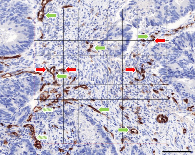
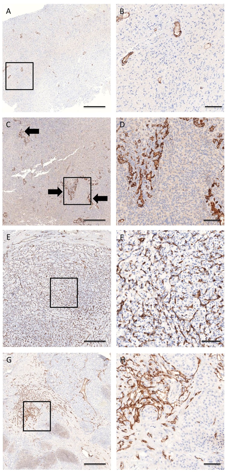
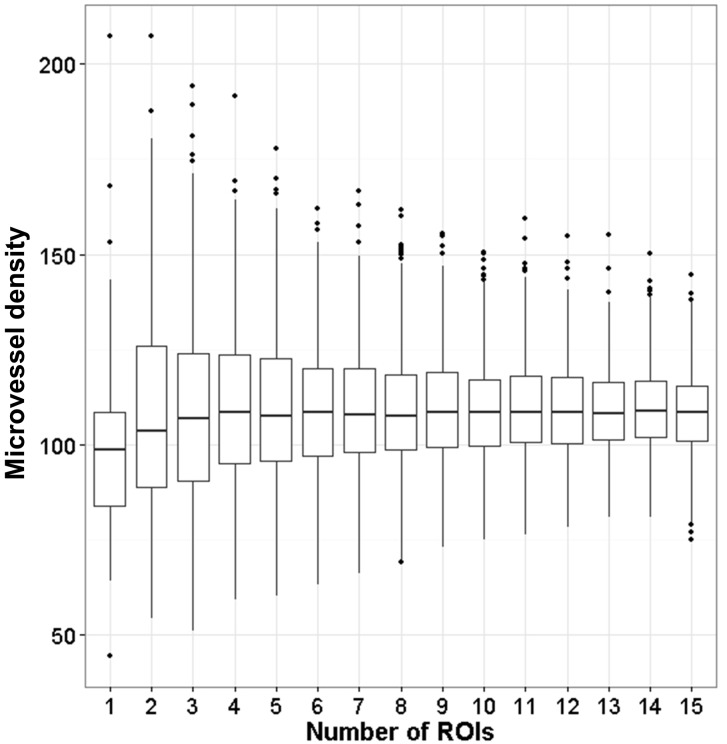
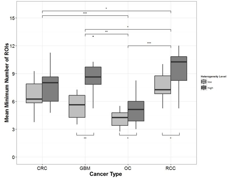
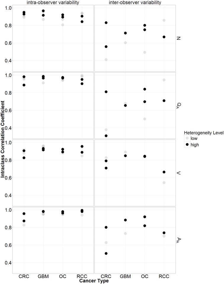
Despite all efforts made to develop predictive biomarkers for antiangiogenic therapies, no unambiguous markers have been identified so far. This is due to among others the lack of standardized tests. This study presents an improved microvessel density quantification method in tumor tissue based on stereological principles and using whole-slide images. Vessels in tissue sections of different cancer types were stained for CD31 by an automated and validated immunohistochemical staining method. The stained slides were digitized with a digital slide scanner. Systematic, uniform, random sampling of the regions of interest on the whole-slide images was performed semi-automatically with the previously published applications AutoTag and AutoSnap. Subsequently, an unbiased counting grid was combined with the images generated with these scripts. Up to six independent observers counted microvessels in up to four cancer types: colorectal carcinoma, glioblastoma multiforme, ovarian carcinoma and renal cell carcinoma. At first, inter-observer variability was found to be unacceptable. However, after a series of consensus training sessions and interim statistical analysis, counting rules were modified and inter-observer concordance improved considerably. Every CD31-positive object was counted, with exclusion of suspected CD31-positive monocytes, macrophages and tumor cells. Furthermore, if interconnected, stained objects were considered a single vessel. Ten regions of interest were sufficient for accurate microvessel density measurements. Intra-observer and inter-observer variability were low (intraclass correlation coefficient > 0.7) if the observers were adequately trained.


Conflict of interest statement

The authors have read the journal's policy and the authors of this manuscript have the following competing interests: Koen Marien, Valerie Croons, Yannick Waumans, Ellen Sluydts, Stefanie De Schepper, and Luc Andries are employees of HistoGeneX NV (Antwerp, Belgium). Mark Kockx is CEO of HistoGeneX NV, which carries out immunohistochemical testing for pharmaceutical companies as part of (pre-)clinical studies that evaluate new anticancer drugs. Mark Kockx and Wim Waelput are consulting pathologists to Pathomation BVBA. The authors have no other relevant affiliations or financial involvements with any organization or entity with a financial interest in or financial conflict with the subject matter or materials discussed in the manuscript apart from those disclosed. This does not alter the authors' adherence to PLOS ONE policies on sharing data and materials.

Figures

Fig 1
Fig 1. Example of a region of interest.
It was captured in Pannoramic Viewer (3DHISTECH, Budapest, Hungary) and combined with a digital 81-points grid in Adobe Photoshop CS4. CD31-stained vessel profiles in the grid were counted as N (green arrow). Vessel profiles that cross the virtually extended left or lower line of the grid were not counted (shaded green arrow). The grid points that hit a CD31-stained vascular profile were counted as V (red arrow). Scale bar represents 100 μm.
Fig 2
Fig 2. Histological heterogeneity of CD31-stained blood vessels in glioblastoma multiforme (a-d) and renal cell carcinoma (e-h).
(a-b) QA = 15 vessels per mm2, AA = 1.56%, (c-d) QA = 77 vessels per mm2, AA = 3.70%, (e-f) QA = 183 vessels per mm2, AA = 13.10%, (g-h) QA = 81 vessels per mm2, AA = 6.17%. Low (a, b, e, f) heterogeneous samples showed a uniform distribution of vessel profiles as compared to high (c, d, g, h) heterogeneous samples. In glioblastoma multiforme, hotspots and garlands (arrows) were more easily recognized in heterogeneous than in homogeneous samples. Scale bar represents 500 μm (a, c, e, g) or 100 μm (b, d, f, h)
Fig 3
Fig 3. Distribution of 1000 bootstrap results.
Here the results for CRC sample 19 is displayed. These were calculated based on the counting by the second observer during the second round of counting. Tukey boxplots were constructed for amounts of regions of interest evaluated. Ten regions are sufficient for accurate microvessel density calculation.
Fig 4
Fig 4. Tukey boxplots illustrating the relationship between the mean minimum number of regions of interest (ROIs) and the topological blood vessel heterogeneity.
This was done for each sample and for every cancer type: colorectal carcinoma (CRC), glioblastoma multiforme (GBM), ovarian carcinoma (OC), and renal cell carcinoma (RCC). If the topological blood vessel heterogeneity of the samples increased (low < high), the minimum number of ROIs on average increased as well. ***p < 0.001, **p < 0.01, *p < 0.05.
Fig 5
Fig 5. Intra- (left) and inter-observer (right) variability for the old counting rules.
This was calculated by the intraclass correlation coefficients (ICC) for the four parameters (V, N, QA, AA). In the first row this is displayed for the number of vessel profiles in a region of interest (N). In the second row this is displayed for the microvessel density (QA). In the third row this is displayed for the number of points in the grid hitting a vessel profile in a region of interest (V). In the last row this is displayed for the areal fraction of vessel profiles (AA). In addition are the ICCs in relation to the heterogeneity level (low or high) and the cancer type (colorectal carcinoma (CRC), glioblastoma multiforme (GBM), ovarian carcinoma (OC), and renal cell carcinoma (RCC)) shown.
Fig 6
Fig 6. Inter-observer variation for the old counting rules between observer 1 (KM) and 2 (VC) for colorectal cancer samples.
This was displayed by Bland-Altman (a, c, e, g) and prediction plots with prediction intervals (two black lines) (b, d, f, h) for the number of vessel profiles (N) (a, b), the microvessel density (QA) (c,d), the number of points in the grid hitting a vessel profile (V) (e, f) and the areal fraction of vessel profiles (AA) (g, h). A systemic bias for N, QA, and AA was present as illustrated by the prediction plots (large distance between the x = y line (black and dashed) and the linear regression line of the measurements (red)).

Similar articles

Cited by

References

    1. Donnem T, Hu J, Ferguson M, Adighibe O, Snell C, Harris AL, et al. Vessel co-option in primary human tumors and metastases: an obstacle to effective anti-angiogenic treatment? Cancer Med. 2013;2: 427–36. 10.1002/cam4.105 - DOI - PMC - PubMed
    1. Hanahan D, Weinberg RA. Hallmarks of cancer: the next generation. Cell. 2011;144: 646–74. 10.1016/j.cell.2011.02.013 - DOI - PubMed
    1. Weidner N. Current pathologic methods for measuring intratumoral microvessel density within breast carcinoma and other solid tumors. Breast Cancer Res Treat. 1995;36: 169–80. - PubMed
    1. Vermeulen PB, Gasparini G, Fox SB, Toi M, Martin L, McCulloch P, et al. Quantification of angiogenesis in solid human tumours: an international consensus on the methodology and criteria of evaluation. Eur J Cancer. 1996;32A: 2474–84. - PubMed
    1. Vermeulen PB, Gasparini G, Fox SB, Colpaert C, Marson LP, Gion M, et al. Second international consensus on the methodology and criteria of evaluation of angiogenesis quantification in solid human tumours. Eur J Cancer. 2002;38: 1564–79. - PubMed

Publication types

MeSH terms

Substances

LinkOut - more resources