

Hypereosinophilic syndrome presenting with multiple organ infiltration and deep venous thrombosis: A case report and literature review
- PMID: 27583887
- PMCID: PMC5008571
- DOI: 10.1097/MD.0000000000004658
Hypereosinophilic syndrome presenting with multiple organ infiltration and deep venous thrombosis: A case report and literature review
Abstract
Background: Hypereosinophilic syndrome (HES) can be fatal, particularly when eosinophils infiltrate vital organs and/or if extensive thrombosis develops. However there are no standard recommendations for the use of anticoagulant therapy of HES in the setting of thrombosis.
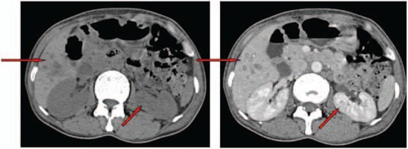
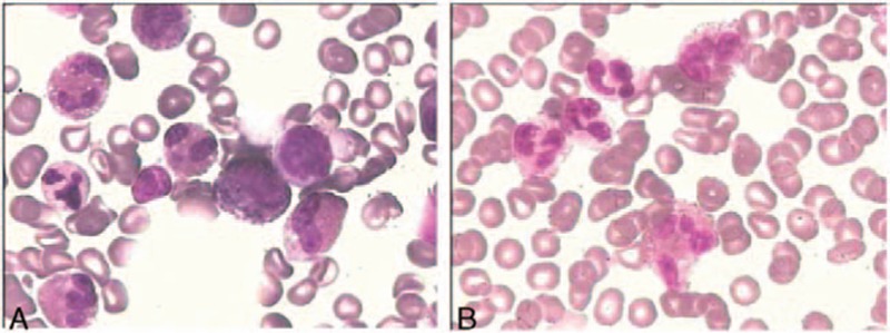
Methods: We herein present a case of a 46-year-old female who presented with marked peripheral eosinophilia with symptoms of multi-organ infiltration and extensive deep venous thrombosis (DVT). In this case, evaluation was carried out before the diagnosis was established, and timely standard-dose corticosteroids combined with a new oral anticoagulant (NOAC) therapy were carried out.
Results: These measures resulted in a rapid response and long-term disease control.
Conclusion: Although there are no data to support which anticoagulant is preferred in this setting, this case indicates that the new oral anticoagulants may play an important role in the treatment of thrombosis in HES.
Conflict of interest statement
The authors have no conflicts of interest to disclose.
Figures


References
-
- Gotlib J. World Health Organization-defined eosinophilic disorders: 2015 update on diagnosis, risk stratification, and management. Am J Hematol 2015; 90:1077–1089. - PubMed
-
- Podjasek JC, Butterfield JH. Mortality in hypereosinophilic syndrome: 19 years of experience at Mayo Clinic with a review of the literature. Leuk Res 2013; 37:392–395. - PubMed
-
- Kahn JE. Hypereosinophilic syndromes. Best Pract Res Clin Rheumatol 2008; 22:863–882. - PubMed
-
- Narayan S, Ezughah F, Standen GR, et al. Idiopathic hypereosinophilic syndrome associated with cutaneous infarction and deep venous thrombosis. Br J Dermatol 2003; 148:817–820. - PubMed
Publication types
MeSH terms
Substances
LinkOut - more resources
Full Text Sources
Other Literature Sources
Medical
