

Nitric oxide released from JS-K induces cell death by mitotic catastrophe as part of necrosis in glioblastoma multiforme
- PMID: 27584787
- PMCID: PMC5059858
- DOI: 10.1038/cddis.2016.254
Nitric oxide released from JS-K induces cell death by mitotic catastrophe as part of necrosis in glioblastoma multiforme
Abstract
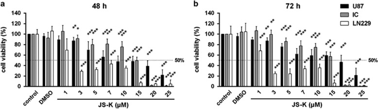
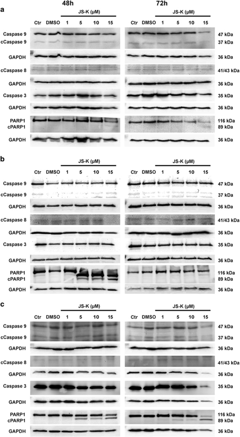
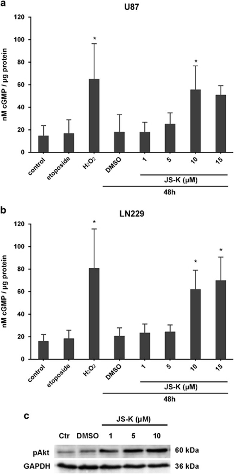
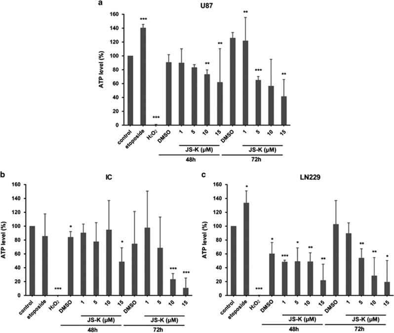
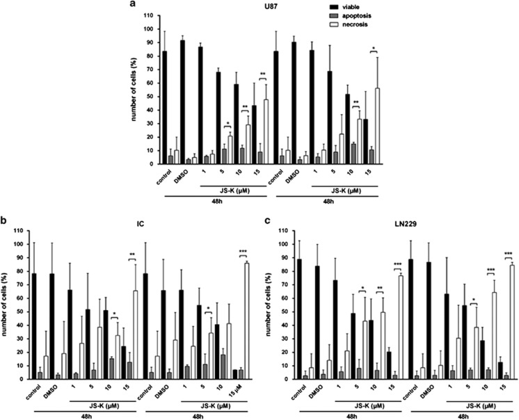
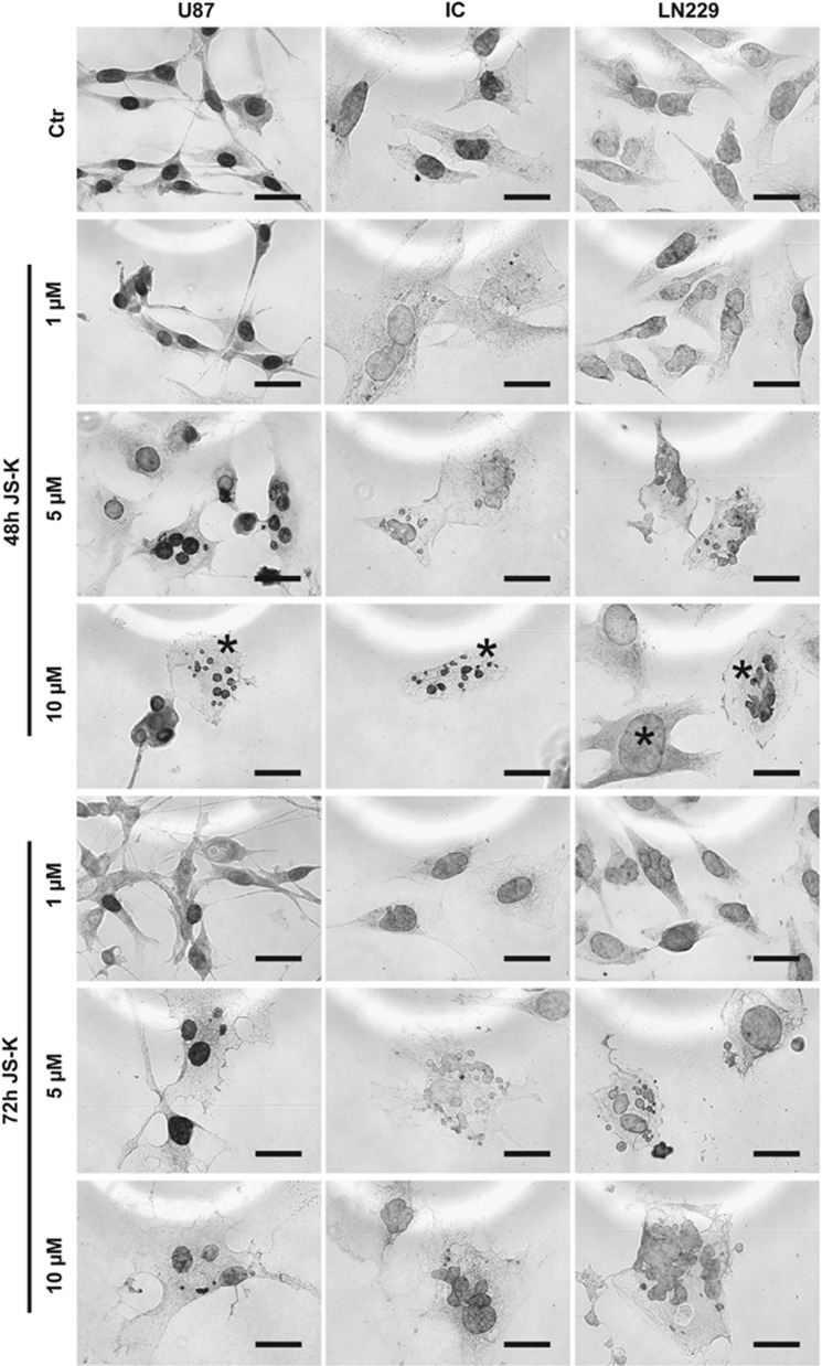
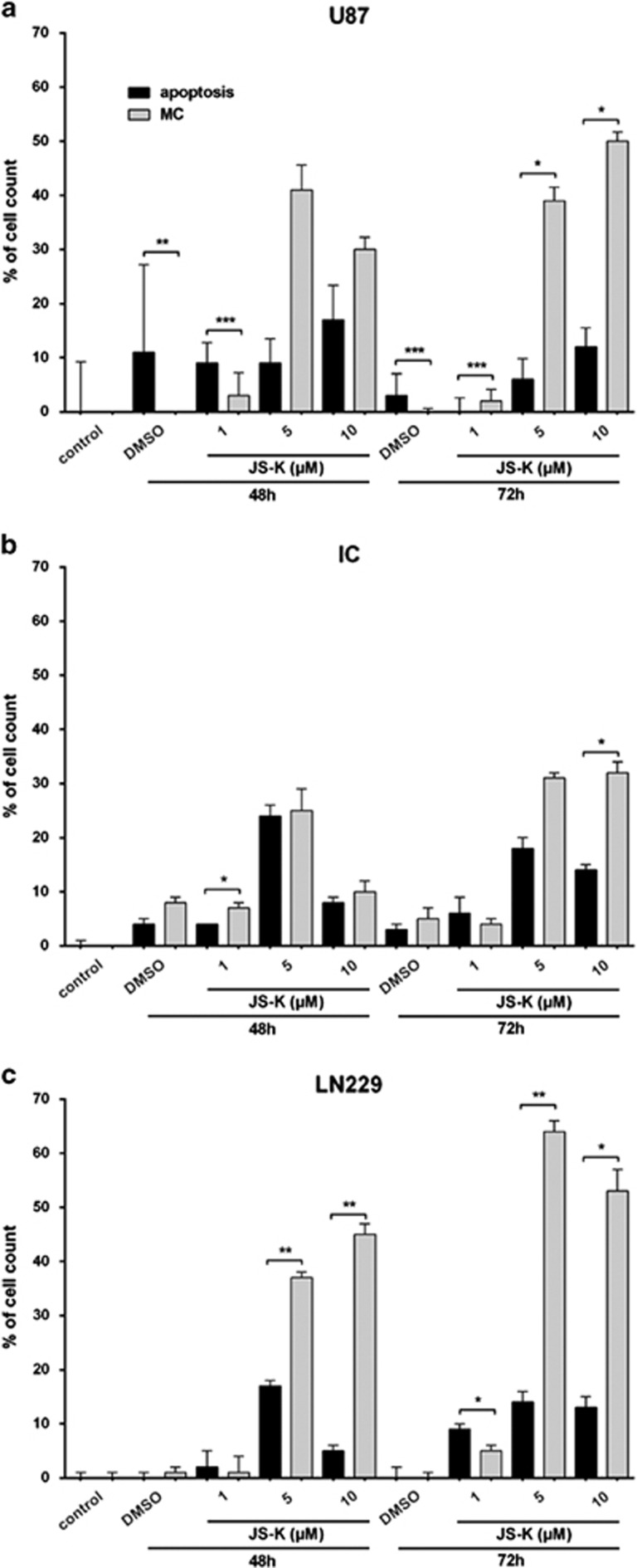
The nitric oxide (NO) donor JS-K is specifically activated by glutathione S-transferases (GSTs) in GST-overexpressing cells. We have shown the induction of cell death in glioblastoma multiforme (GBM) cells at high JS-K doses but the mechanism remains unclear. The aim of this study was to determine whether NO-induced cell death is triggered by induction of apoptotic or necrotic pathways. For the first time, we demonstrate that NO induces cell death via mitotic catastrophe (MC) with non-apoptotic mechanisms in GBM cells. Moreover, the level of morphological changes indicating MC correlates with increased necrosis. Therefore, we conclude that MC is the main mechanism by which GBM cells undergo cell death after treatment with JS-K associated with necrosis rather than apoptosis. In addition, we show that PARP1 is not an exclusive marker for late apoptosis but is also involved in MC. Activating an alternative way of cell death can be useful for the multimodal cancer therapy of GBM known for its strong anti-apoptotic mechanisms and drug resistance.
Figures
Similar articles
-
Cyclooxygenase (COX) Inhibition by Acetyl Salicylic Acid (ASA) Enhances Antitumor Effects of Nitric Oxide in Glioblastoma In Vitro.Mol Neurobiol. 2019 Sep;56(9):6046-6055. doi: 10.1007/s12035-019-1513-6. Epub 2019 Feb 4. Mol Neurobiol. 2019. PMID: 30715649
-
JS-K, a glutathione S-transferase-activated nitric oxide donor with antineoplastic activity in malignant gliomas.Neurosurgery. 2012 Feb;70(2):497-510; discussion 510. doi: 10.1227/NEU.0b013e31823209cf. Neurosurgery. 2012. PMID: 21849924 Free PMC article.
-
JS-K, a nitric oxide donor, induces autophagy as a complementary mechanism inhibiting ovarian cancer.BMC Cancer. 2019 Jul 1;19(1):645. doi: 10.1186/s12885-019-5619-z. BMC Cancer. 2019. PMID: 31262254 Free PMC article.
-
Biology of Glioblastoma Multiforme-Exploration of Mitotic Catastrophe as a Potential Treatment Modality.Int J Mol Sci. 2020 Jul 27;21(15):5324. doi: 10.3390/ijms21155324. Int J Mol Sci. 2020. PMID: 32727112 Free PMC article. Review.
-
Molecular definitions of cell death subroutines: recommendations of the Nomenclature Committee on Cell Death 2012.Cell Death Differ. 2012 Jan;19(1):107-20. doi: 10.1038/cdd.2011.96. Epub 2011 Jul 15. Cell Death Differ. 2012. PMID: 21760595 Free PMC article. Review.
Cited by
-
Inducible nitric oxide synthase-derived extracellular nitric oxide flux regulates proinflammatory responses at the single cell level.Redox Biol. 2020 Jan;28:101354. doi: 10.1016/j.redox.2019.101354. Epub 2019 Nov 1. Redox Biol. 2020. PMID: 31683257 Free PMC article.
-
JS-K induces reactive oxygen species-dependent anti-cancer effects by targeting mitochondria respiratory chain complexes in gastric cancer.J Cell Mol Med. 2019 Apr;23(4):2489-2504. doi: 10.1111/jcmm.14122. Epub 2019 Jan 22. J Cell Mol Med. 2019. PMID: 30672108 Free PMC article.
-
Cyclooxygenase (COX) Inhibition by Acetyl Salicylic Acid (ASA) Enhances Antitumor Effects of Nitric Oxide in Glioblastoma In Vitro.Mol Neurobiol. 2019 Sep;56(9):6046-6055. doi: 10.1007/s12035-019-1513-6. Epub 2019 Feb 4. Mol Neurobiol. 2019. PMID: 30715649
-
Metformin in combination with JS-K inhibits growth of renal cell carcinoma cells via reactive oxygen species activation and inducing DNA breaks.J Cancer. 2020 Mar 31;11(13):3701-3712. doi: 10.7150/jca.36372. eCollection 2020. J Cancer. 2020. PMID: 32328174 Free PMC article.
-
ATF3 reduces migration capacity by regulation of matrix metalloproteinases via NFκB and STAT3 inhibition in glioblastoma.Cell Death Discov. 2017 Feb 27;3:17006. doi: 10.1038/cddiscovery.2017.6. eCollection 2017. Cell Death Discov. 2017. PMID: 28250971 Free PMC article.
References
-
- Contestabile A, Ciani E. Role of nitric oxide in the regulation of neuronal proliferation, survival and differentiation. Neurochem Int 2004; 45: 903–914. - PubMed
MeSH terms
Substances
LinkOut - more resources
Full Text Sources
Other Literature Sources
Research Materials
Miscellaneous
