. 2016 Sep 1;7(9):e2353.
doi: 10.1038/cddis.2016.253.

Epstein-Barr virus-encoded miR-BART6-3p inhibits cancer cell metastasis and invasion by targeting long non-coding RNA LOC553103

Affiliations

Epstein-Barr virus-encoded miR-BART6-3p inhibits cancer cell metastasis and invasion by targeting long non-coding RNA LOC553103

Baoyu He et al. Cell Death Dis. .

Abstract

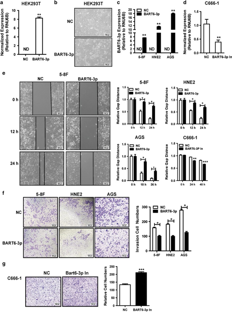
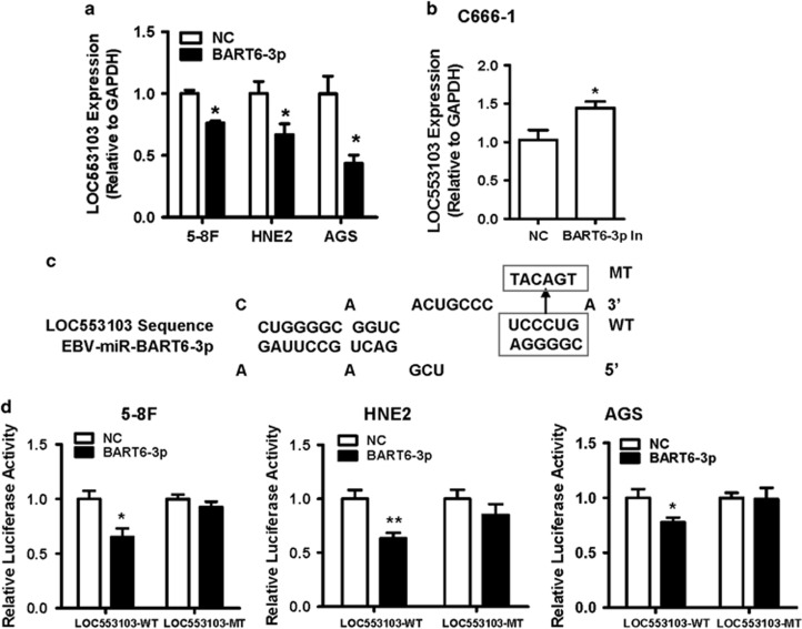
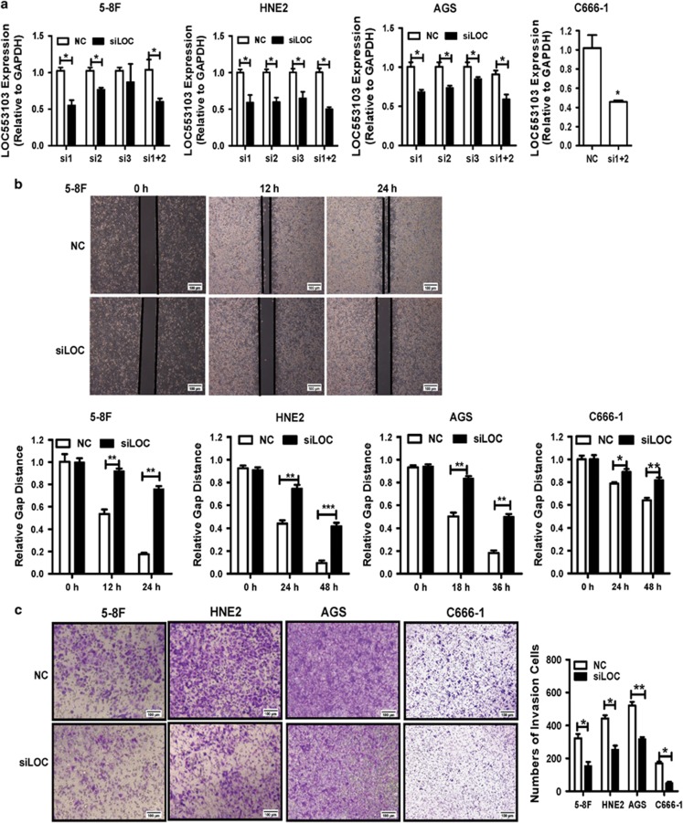
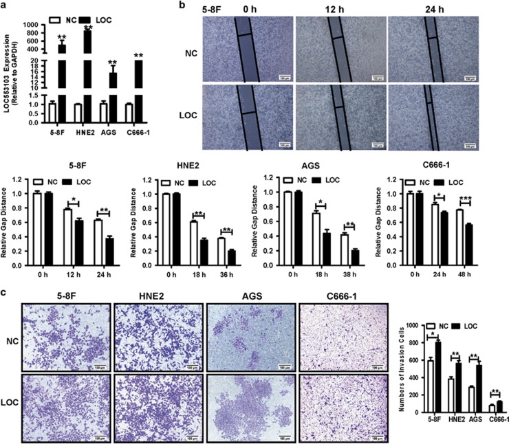
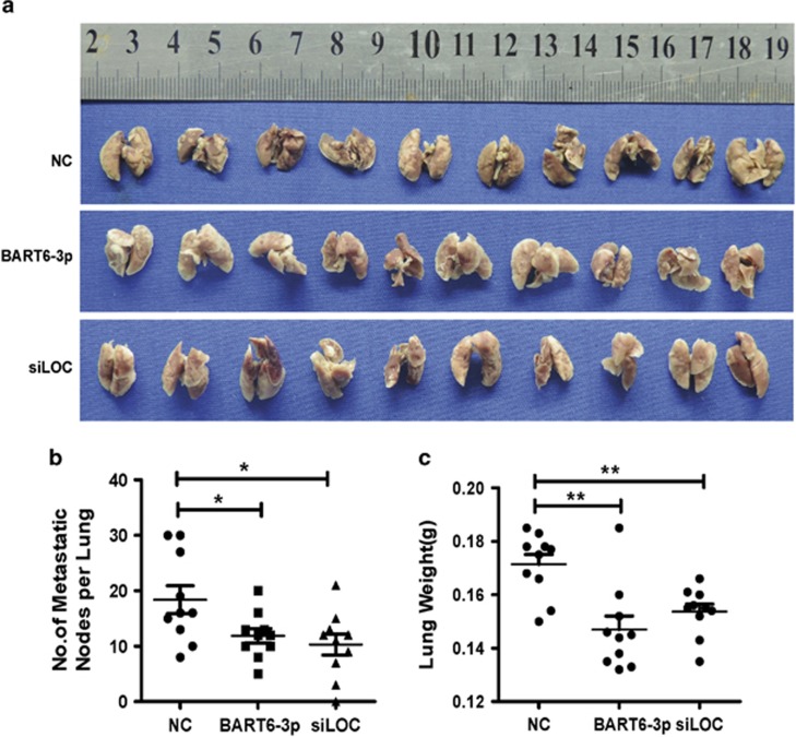
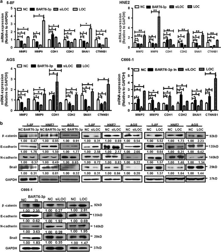
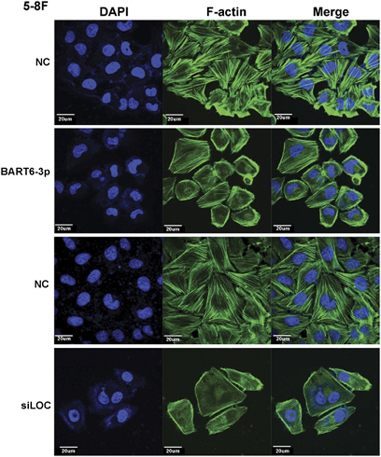
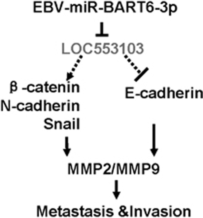
Epstein-Barr virus (EBV) infection is causatively related to a variety of human cancers, including nasopharyngeal carcinoma (NPC) and gastric cancer (GC). EBV encodes 44 mature miRNAs, a number of which have been proven to promote carcinogenesis by targeting host genes or self-viral genes. However, in this study, we found that an EBV-encoded microRNA, termed EBV-miR-BART6-3p, inhibited EBV-associated cancer cell migration and invasion including NPC and GC by reversing the epithelial-mesenchymal transition (EMT) process. Using microarray analysis, we identified and validated that a novel long non-coding RNA (lncRNA) LOC553103 was downregulated by EBV-miR-BART6-3p, and LOC553103 knockdown by specific siRNAs phenocopied the effect of EBV-miR-BART6-3p, while LOC553103 overexpression promoted cancer cell migration and invasion to facilitate EMT. In conclusion, we determined that EBV-miR-BART6-3p, a microRNA encoded by oncogenic EBV, inhibited EBV-associated cancer cell migration and invasion by targeting and downregulating a novel lncRNA LOC553103. Thus, our study presents an unreported mechanism underlying EBV infection in EBV-associated cancer carcinogenesis, and provides a potential novel diagnosis and treatment biomarker for NPC and other EBV-related cancers.


Figures

Figure 1
Figure 1
EBV-miR-BART6-3p mimics suppresses tumor cell migration and invasion in vitro. The bars indicate mean±S.D. (n=3). *P<0.05; **P<0.01; ***P<0.001. (a) Exogenous EBV-miR-BART6-3p expression was detected by real-time PCR after transfection of HEK293T cells with EBV-miR-BART6-3p mimics (BART6-3p) or negative control (NC). ND, not detectable. (b) Morphological alterations in HEK293T cells upon BART6-3p mimic transfection, as assessed by phase contrast microscopy ( × 100). (c) Expression of exogenous EBV-miR-BART6-3p was detected in HNE2, 5-8 F and AGS cells after transfection with EBV-miR-BART6-3p mimics (BART6-3p) or negative control (NC) in EBV-negative NPC cell lines (HNE2 and 5-8 F) or gastric cell line (AGS). (d) Expression of exogenous EBV-miR-BART6-3p was detected in C666-1 cells after transfection with EBV-miR-BART6-3p inhibitor (BART6-3p In) or negative control (NC) in EBV-positive cell line (C666-1). (e) EBV-miR-BART6-3p mimics inhibited 5-8 F, HNE2, and AGS cell migration. EBV-miR-BART6-3p inhibitors accelerated C666-1 cell migration. Cells were grown and transfected with EBV-miR-BART6-3p mimics, EBV-miR-BART6-3p inhibitors or a negative control and subjected to wound healing assays. The left panel shows the representative results of 5-8 F, and the right panel summarizes the relative width ratio of the wound healing gap from three independent experiments. (f) EBV-miR-BART6-3p mimics inhibited tumor cell invasion as measured by transwell matrigel penetration assay. 5-8 F, HNE2 and AGS cells were grown and transfected with EBV-miR-BART6-3p mimics or negative control for 36 h and were subjected to a matrigel invasion assay (left panel). The right graph summarizes the data from three independent experiments. (g) EBV-miR-BART6-3p inhibitors accelerated C666-1 cell invasion as measured by transwell matrigel penetration assay. C666-1 cells were grown and transfected with EBV-miR-BART6-3p inhibitors or negative control for 48 h and were subjected to a matrigel invasion assay (left panel). The right graph summarizes the data from three independent experiments
Figure 2
Figure 2
LOC553103 is a direct EBV-miR-BART6-3p target. The bars indicate mean±S.D. (n=3). *P<0.05; **P<0.01; ***P<0.001. (a) LOC553103 expression was inhibited by EBV-miR-BART6-3p mimics in 5-8 F, HNE2 and AGS cells. (b) LOC553103 expression was induced by EBV-miR-BART6-3p inhibitors in C666-1 cells. (c) The location of the possible seed-matched sites for EBV-miR-BART6-3p on LOC553103 sequence and the sites changed to produce mutated forms of LOC553103. The EBV-miR-BART6-3p seed region had a complementary binding site in the LOC553103 sequence. For the mutated (Mut) form, the potential binding nucleotides were replaced to disrupt the complementarities. (d) 5-8 F, HNE2 and AGS cells were transiently co-transfected with EBV-miR-BART6-3p (BART6-3p) or negative control (NC) as well as pRL-TK and luciferase reporters containing either wild type (LOC553103-WT) or mutated (LOC553103-mut) EBV-miR-BART6-3p-binding site. The cells were analyzed for luciferase activity after 48 h. The experiments were repeated three times, and the error bars denote the mean. EBV-miR-BART6-3p mimics attenuated LOC553103-WT luciferase activity compared with the LOC553103-mutant
Figure 3
Figure 3
LOC553103-siRNA knockdown inhibits cancer cell migration and invasion in vitro. The graph summarizes the data from three independent experiments. The bars indicate mean±S.D. (n=3). *P<0.05; **P<0.01; ***P<0.001. (a) The efficiency of LOC553103 knockdown was measured by examining LOC553103 expression in 5-8 F, HNE2 and AGS cells 48 h after transfection with the siLOC553103 or the scrambled control were measured by real-time PCR. GAPDH was used as an internal control for real-time PCR. All three siRNA sequences (si1, si2 and si3) decreased LOC553103 expression in 5-8 F, HNE2 and AGS cells, but siRNA1 and siRNA2 had better effect than siRNA3, so the mixture of siRNA1 and siRNA2 was used for LOC553103 knockdown in subsequent experiments. The efficiency of LOC553103 knockdown was also measured by examining LOC553103 expression in C666-1 cells transfected with the mixture of siRNA1 and siRNA2. (b) LOC553103 knockdown inhibited 5-8 F, HNE2, AGS and C666-1 cell migration. Cells were grown and transfected with LOC553103 siRNA (siLOC) or scrambled negative control (NC) and then subjected to the wound healing assay. The upper panel shows the representative results of 5-8 F, and the lower panel summarizes the relative width ratio of the wound healing gap from three independent experiments. (c) LOC553103 knockdown inhibited tumor cell invasion as measured by transwell matrigel penetration assay. 5-8 F, HNE2, AGS and C666-1 cells were grown and transfected with LOC553103 siRNAs (siLOC) or scrambled negative control (NC) and then subjected to a matrigel invasion assay
Figure 4
Figure 4
LOC553103 overexpression promotes cancer cell migration and invasion in vitro. The graph summarizes the data from three independent experiments. The bars indicate mean±S.D. (n=3). *P<0.05; **P<0.01; ***P<0.001. (a) LOC553103 expression levels in 5-8 F, HNE2, AGS and C666-1 cells were measured by real-time PCR 48 h after transfection with the LOC553103 overexpression vector (LOC) or negative control empty vector (NC). GAPDH was used as an internal control. (b) LOC553103 induced 5-8 F, HNE2, AGS and C666-1 cell migration. Cells transfected with LOC553103 overexpression vector (LOC) or negative control empty vector (NC) were subjected to the wound healing assay. The upper panel shows representative results of 5-8 F, and the lower panel summarizes the relative width ratio of the wound healing gap from three independent experiments. (c) LOC553103 promoted tumor cell invasion as measured by transwell matrigel penetration assay. 5-8 F, HNE2, AGS and C666-1 cells were transfected with LOC553103 overexpression vector (LOC) or the empty vector (NC) and then subjected to a matrigel invasion assay (left panel)
Figure 5
Figure 5
EBV-miR-BART6-3p overexpression or LOC553103 knockdown inhibits metastasis in nude mice. (a) A total 30 BALB/c nude mice aged 4 weeks were divided into three groups (10 mice per group), and each mouse was injected with 1 × 10⁶ 5-8 F cells transfected with EBV-miR-BART6-3p mimics (BART6-3p), LOC553103 siRNAs (siLOC) or scrambled control sequence (NC) into the tail vein. After 2 months, the mice were killed, and their lungs were removed for assessment of the metastasized tumor nodules. (b) Numbers of metastasized tumor nodules in the lung per mouse were counted. (c) Mouse lung weights were scaled and reflected the decreased metastasized tumor foci size
Figure 6
Figure 6
EBV-miR-BART6-3p and LOC55310 regulate EMT and metastasis-related gene expression. (a) 5-8 F, HNE2, AGS or C666-1 cells were transfected with EBV-miR-BART6-3p mimics (BART6-3p) or EBV-miR-BART6-3p inhibitors (BART6-3p In), LOC553103 siRNAs (siLOC) or LOC553103 overexpression vector. Forty-eight hours after transfection, cells were harvested and mRNA expression levels of metastasis-related genes MMP2, MMP9, EMT-related genes CDH1 (encoding E-cadherin protein), CDH2 (encoding N-cadherin) SNAL1 and CTTNB1 (encoding β-catenin) were measured by real-time PCR. (b) Important EMT markers β-catenin, E-cadherin, N-cadherin and Snail protein expression were measured by western blot 48 h after transfection with EBV-miR-BART6-3p mimics (BART6-3p) or EBV-miR-BART6-3p inhibitors (BART6-3p In), LOC553103 siRNAs (siLOC) or LOC553103 overexpression vector in 5-8 F, HNE2, AGS or C666-1 cells
Figure 7
Figure 7
Transfection of EBV-miR-BART6-3p or LOC55310 siRNAs destroys stress fiber integrity in 5-8 F cells. 5-8 F cells were transfected with EBV-miR-BART6-3p (BART6-3p) mimics, LOC553103 siRNAs (siLOC) or scrambled control (NC), and 48 h after transfection, cells were fixed and stained for F-actin by phalloidin (green), and 4′,6-diamidino-2-phenylindole was used to stain nuclei (blue). A clear deficiency in stress fiber formation was observed in BART6-3p mimic and siLOC553103-transfected 5-8 F cells. Images were acquired at × 400. Scale bar=20 μm
Figure 8
Figure 8
A schematic model of EBV-miR-BART6-3p functioning on cancer invasion and migration and stress fiber integrity through inhibition of its target LOC553103

Similar articles

Cited by

References

    1. Young LS, Rickinson AB. Epstein-Barr virus: 40 years on. Nat Rev Cancer 2004; 4: 757–768. - PubMed
    1. Young LS, Murray PG. Epstein-Barr virus and oncogenesis: from latent genes to tumours. Oncogene 2003; 22: 5108–5121. - PubMed
    1. Nishikawa J, Yoshiyama H, Iizasa H, Kanehiro Y, Nakamura M, Nishimura J et al. Epstein-barr virus in gastric carcinoma. Cancers 2014; 6: 2259–2274. - PMC - PubMed
    1. He JR, Tang LY, Yu DD, Su FX, Song EW, Lin Y et al. Epstein-Barr virus and breast cancer: serological study in a high-incidence area of nasopharyngeal carcinoma. Cancer Lett 2011; 309: 128–136. - PubMed
    1. Rowe M, Fitzsimmons L, Bell AI. Epstein-Barr virus and Burkitt lymphoma. Chin J Cancer 2014; 33: 609–619. - PMC - PubMed

MeSH terms