. 2016 Sep 1;7(9):e2354.
doi: 10.1038/cddis.2016.250.

HSV-1-induced activation of NF-κB protects U937 monocytic cells against both virus replication and apoptosis

Affiliations

HSV-1-induced activation of NF-κB protects U937 monocytic cells against both virus replication and apoptosis

Francesca Marino-Merlo et al. Cell Death Dis. .

Abstract

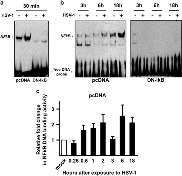
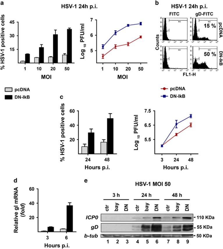
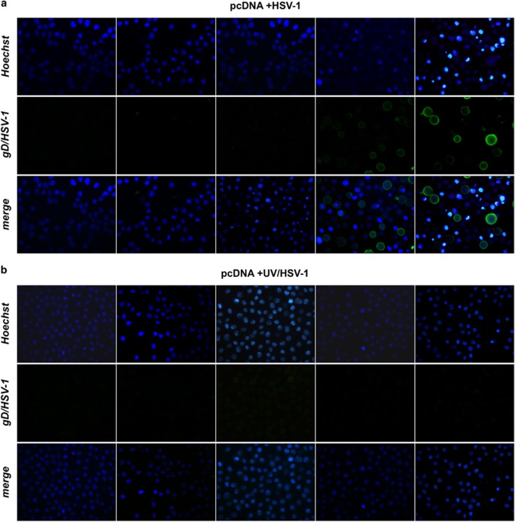
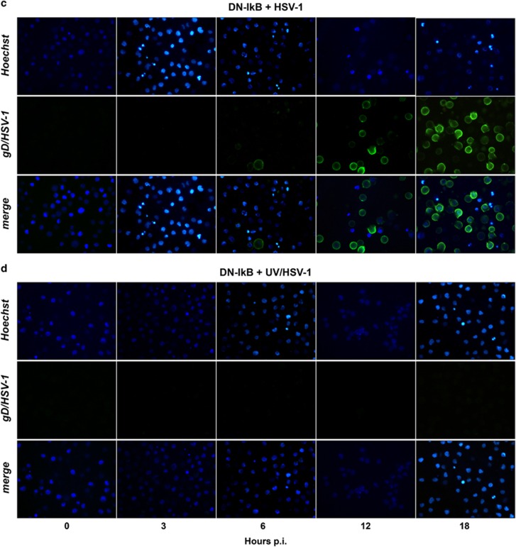
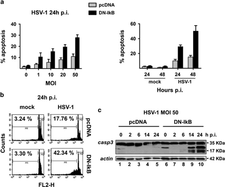
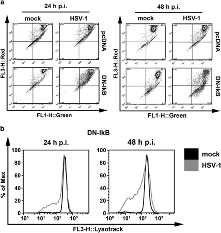
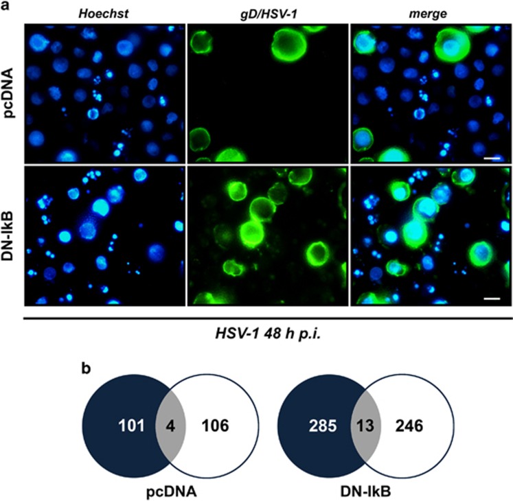
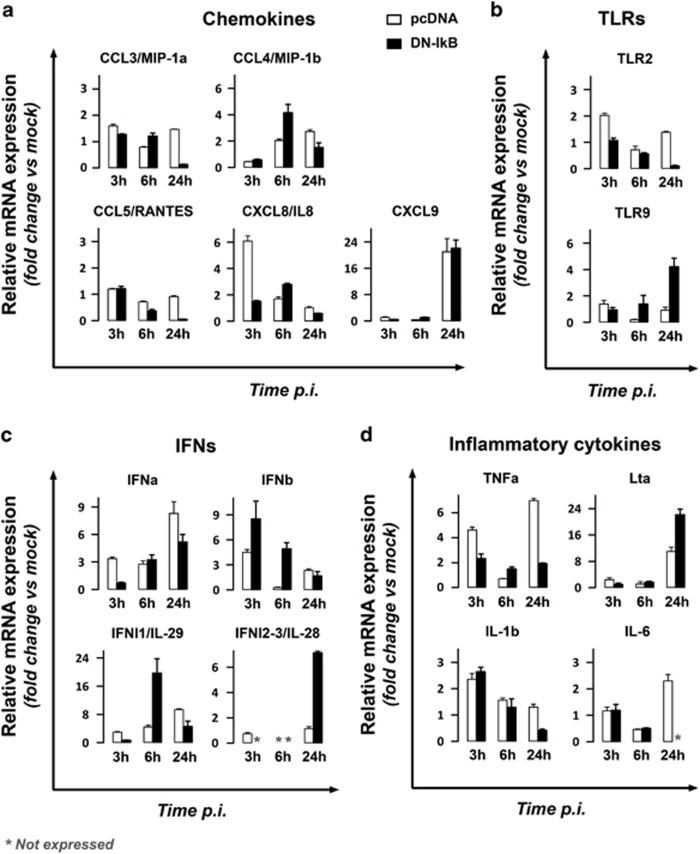
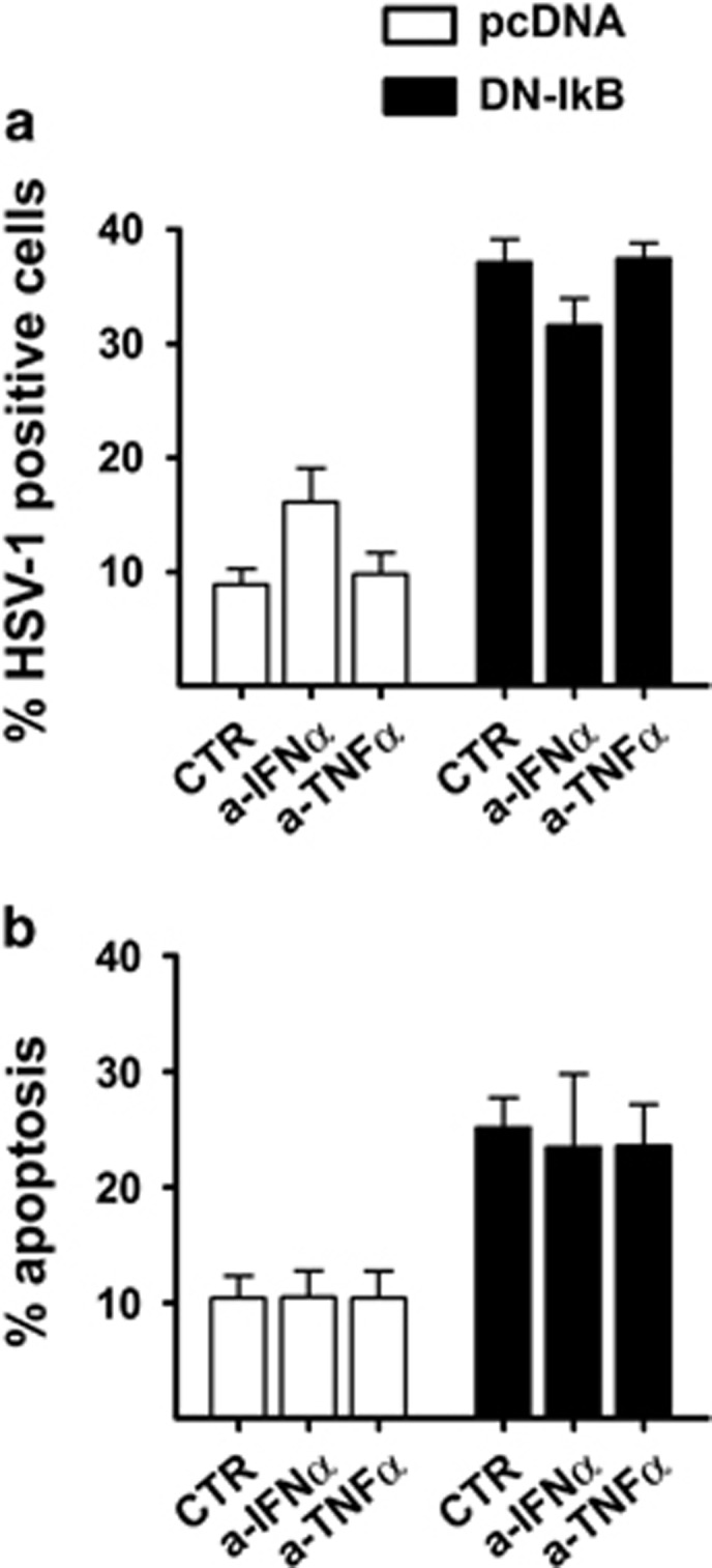
The transcription factor nuclear factor-kappa B (NF-κB) is a crucial player of the antiviral innate response. Intriguingly, however, NF-κB activation is assumed to favour herpes simplex virus (HSV) infection rather than restrict it. Apoptosis, a form of innate response to viruses, is completely inhibited by HSV in fully permissive cells, but not in cells incapable to fully sustain HSV replication, such as immunocompetent cells. To resolve the intricate interplay among NF-κB signalling, apoptosis and permissiveness to HSV-1 in monocytic cells, we utilized U937 monocytic cells in which NF-κB activation was inhibited by expressing a dominant-negative IκBα. Surprisingly, viral production was increased in monocytic cells in which NF-κB was inhibited. Moreover, inhibition of NF-κB led to increased apoptosis following HSV-1 infection, associated with lysosomal membrane permeabilization. High expression of late viral proteins and induction of apoptosis occurred in distinct cells. Transcriptional analysis of known innate response genes by real-time quantitative reverse transcription-PCR excluded a contribution of the assayed genes to the observed phenomena. Thus, in monocytic cells NF-κB activation simultaneously serves as an innate process to restrict viral replication as well as a mechanism to limit the damage of an excessive apoptotic response to HSV-1 infection. This finding may clarify mechanisms controlling HSV-1 infection in monocytic cells.


Figures

Figure 1
Figure 1
NF-κB DNA binding activity by EMSA in U937 monocytic cells infected by HSV-1. U937 cells stably transfected with an empty control vector (pcDNA) or with a dominant negative (DN) mutant of murine IκBα (DN-IkB), were mock infected (−) or infected (+) with 50 MOIs of HSV-1. At 30 min or 3, 6 and 18 h after the first exposure to virus, nuclear extracts were prepared and NF-κB activation was measured by EMSA. pcDNA and DN-IkB samples collected at 30 min were run in the same gel (a), while pcDNA and DN-IkB samples collected at the other time points were run in separate gels (b). The position of the NF-κB DNA is indicated. EMSA gels from one representative experiment of the three performed are shown. (c) Quantitative analysis of the data shown in (a) and (b) using densitometry image analysis of the EMSA gels. Results are expressed as fold change in band intensities from HSV-1- versus mock-infected samples at the same time point. Data represent the mean values±s.d. of three separate experiments for each time course. Multiple comparisons by Bonferroni's post-hoc ANOVA test gave the following results for time 0.25 h versus other times: versus 1 h, P=0.044; versus 2 h and versus 18 h, P=0.002; versus 6 h, P<0.001; versus all other times, not significant. No appreciable optical densitometry value was detected in EMSA gels from parallel time course experiments in U937-DN-IκB cells (data not shown)
Figure 2
Figure 2
Rate of HSV-1 replication in the presence or in the absence of NF-κB activation in U937 cells. U937-pcDNA and U937-DN-IκB cells were assayed for virus replication by different techniques. (a) Cells were infected with different MOIs (1-50) of HSV-1 and analysed at 24 h p.i. by indirect immunofluorescence (left panel) using HSV-1 gD-specific antibody or by plaque-forming assay on Vero cells (right panel). Percentage of gD-HSV-1 positive cells was determined by counting 10 different fields under fluorescence microscope. Viral titres are expressed as Log10 PFU/ml in Vero cells of total virus yield from cells infected at the indicated MOI and are depicted in a logarithmic scale (right panel). Data in the left and right panels represent mean values±s.d. from three independent experiments. Left panel, multiple comparisons by Bonferroni's post-hoc ANOVA test gave the following results: pc-DNA versus corresponding DN-IkB groups at all MOI assayed, P<0.001; among the pcDNA groups, MOI 1 versus MOI 50, P=0.002, all other comparisons, not significant; among the DN-IkB groups, MOI 1 versus MOI 10, P=0.001, MOI 20 versus MOI 50, P=0.008, all other comparisons, P<0.001. Right panel, pcDNA versus corresponding DN-IkB groups at all MOI assayed, P<0.001; among the pcDNA groups, MOI 10 versus MOI 20, not significant, MOI 20 versus MOI 50, P=0.002, all other comparisons, P<0.001; among the DN-IkB groups, MOI 10 versus MOI 20, P=0.001, MOI 20 versus MOI 50, not significant, all other comparisons, P<0.001. (b) Cells were infected with HSV-1 at an MOI of 50 and analysed at 24 h p.i. for viral protein expression by flow cytometry using HSV-1 gD-specific antibody. (c) Time dependency of virus replication (MOI 50) assayed as in (a) by immunofluorescence (left panel) or plaque-forming assay (right panel). Data represent the mean values±s.d. from four independent experiments. Left panel, multiple comparisons by Bonferroni's post-hoc ANOVA test gave the following results: pcDNA versus corresponding DN-IkB at all times assayed, P<0.001; t24 versus t48, pcDNA not significant, DN-IkB P<0.001. Right panel, pcDNA versus corresponding DN-IkB, t3 not significant, all other comparisons, P<0.001; among pcDNA and DN-IκB groups at all times assayed, P<0.001. (d) qPCR analysis of mRNA levels for HSV-1 glycoprotein I in pcDNA cells infected at an MOI of 50 at the indicated times. Data obtained from three independent experiments performed in duplicate were normalized to 18 S rRNA. Values are expressed as mean±s.d. fold changes, relative to gI mRNA levels at 3 h p.i. (the lowest detected value). Multiple comparisons by Bonferroni's post-hoc ANOVA test gave the following results: t6 versus all other groups, P<0.001; all other comparisons, not significant. (e) Western blot analysis showing the effects of pharmacological (bay lanes 2, 5 and 8) and biochemical (lanes 3, 6 and 9) inhibition of NF-κB activity on expression of IE ICP0 and late gD viral proteins. Whole cell lysates were prepared from HSV-infected (50 MOIs) U937-DN-IκB cells and pcDNA cells, pretreated (bay) or not (ctr) with 1 μM BAY-117085 for 16 h. β-tubulin was used as loading control
Figure 3
Figure 3
Immunofluorescence microscopy analysis of time-dependent expression of gD-HSV-1 in U937 cells infected with HSV-1 inactivated or not with UV irradiation. (a) U937-pcDNA cells were infected with intact HSV-1 (50 PFU/cell). At the end of 1 h adsorption period, the virus inoculum was removed, the cells were washed, fresh medium was added and samples for the first time point were collected and stained (time 0 p.i.). Residual cells were incubated at 37 °C until samples were collected at the indicated hours p.i. Images were captured using fluorescent filters optimized for detecting Hoechst-stained nuclei (upper line), surface gD-expressing cells (middle line) or merged (lower line). (b) Parallel infection performed as described in (a) on U937-pcDNA cells, but using UV-inactivated HSV-1. (c) Parallel infection performed as described in (a) but using U937-DN-IκB cells as target cells. (d) Parallel infection performed as described in (b) but using U937-DN-IκB cells as target cells
Figure 3
Figure 3
Immunofluorescence microscopy analysis of time-dependent expression of gD-HSV-1 in U937 cells infected with HSV-1 inactivated or not with UV irradiation. (a) U937-pcDNA cells were infected with intact HSV-1 (50 PFU/cell). At the end of 1 h adsorption period, the virus inoculum was removed, the cells were washed, fresh medium was added and samples for the first time point were collected and stained (time 0 p.i.). Residual cells were incubated at 37 °C until samples were collected at the indicated hours p.i. Images were captured using fluorescent filters optimized for detecting Hoechst-stained nuclei (upper line), surface gD-expressing cells (middle line) or merged (lower line). (b) Parallel infection performed as described in (a) on U937-pcDNA cells, but using UV-inactivated HSV-1. (c) Parallel infection performed as described in (a) but using U937-DN-IκB cells as target cells. (d) Parallel infection performed as described in (b) but using U937-DN-IκB cells as target cells
Figure 4
Figure 4
Extent of apoptosis induced by HSV-1 in U937 cells. (a) MOI-dependency (left panel) and time-dependency (right panel) of apoptosis as detected by cellular (apoptotic bodies) or nuclear morphology following Hoechst 33342 staining (chromatin condensation, nuclear fragmentation) in HSV-1 infected cells. Apoptotic cells/nuclei were counted in 10 high-power fields for each sample. Data represent mean values±s.d. from three independent experiments. Left panel, multiple comparisons by Bonferroni's post-hoc ANOVA test gave the following results: pc-DNA versus corresponding DN-IkB groups, MOI 0 not significant, MOI 1 P=0.002, all other MOI P<0.001; among the pcDNA groups, MOI 0 versus MOI 20, P=0.003, MOI 0 versus MOI 50, P= 0.001, MOI 10 versus MOI 50, P=0.024, all other comparisons, not significant; among DN-IkB groups, MOI 1 versus MOI 10, P=0.041, MOI 10 versus MOI 20, not significant, all other comparisons, P<0.001. Right panel, pc-DNA versus corresponding DN-IkB groups at all times assayed, mock groups not significant, HSV-1 groups P<0.001; among the pcDNA groups, mock 24 versus mock 48, not significant, mock 24 versus HSV-1 24, P=0.008, mock 24 versus HSV-1 24, mock 24 versus HSV-1 48, P<0.001, mock 48 versus HSV-1 24, P=0.001, mock 48 versus HSV-1 48, P<0.001, HSV-1 24 versus HSV-1 48, not significant; among the DN-IκB groups, mock 24 versus mock 48, not significant, all other comparisons, P< 0.001. (b) Cell death quantitation by FACS analysis using propidium iodide-stained nuclei. U937-pcDNA and U937-DN-IκB cells were mock-infected or infected with 50 MOI of HSV-1and collected after 24 h for sample preparation. M1 and M2 markers indicate the boundaries between the hypodiploid and the diploid nuclei arbitrarily set on the mock-infected samples and maintained for the infected samples. The numbers represent the percentages of hypodiploid nuclei. (c) Western blot analysis of caspase-3 cleavage in HSV-1 infected cells. Whole cell lysates were prepared from both cell lines at the indicated times after infection with 50 MOI HSV-1. Antibodies for procaspase-3 (32 kDa) and the active cleaved form of caspase-3 (20–17 kDa) were used for immunoblot analysis and anti-actin as loading control
Figure 5
Figure 5
Extent of lysosomal membrane permeability after HSV-1 infection of U937-pcDNA and U937-DN-IκB cells. Quantitative, FACS analysis of mock-infected (mock) or HSV-1-infected (HSV-1) cells (MOI 50 PFU cell−1), following staining with 10 μM acridine orange (a) or 75 nM LysoTracker Red DND-99 (b) at 24 h and 48 h p.i. Dot plots (a) and histograms (b) show a greater reduction of red fluorescence in the lysosomes (FL3-H) concomitant with an increase in green fluorescence in the cytosol (FL1-H) in HSV-1-infected U937-DN-IκB cells
Figure 6
Figure 6
Simultaneous detection of apoptotic and gD/HSV-1-positive cells by immunofluorescence analysis of HSV-1-infected U937-pcDNA and U937-DN-IκB cells. (a) Representative microscopy images captured at 48 h p.i., using fluorescent filters optimized for Hoechst-stained nuclei (Hoechst, blue fluorescence, left panel) and surface gD-expressing cells following staining with a mouse anti-gD antibody and an appropriate secondary fluorescein isothiocyanate-conjugated antibody (gD/HSV-1, green fluorescence, middle panel). Images obtained from the same field with different filters were merged (merge, right panel). Fragmented nuclei typical for apoptosis can be observed. Original magnification (630 × ). Scale bar 10 μM. (b)Venn diagrams showing the overlap (grey) of apoptotic (black) and gD (white) positive cells for the same fields of a total of 958 counted U937-pcDNA and a total of 949 counted U937-DN-IκB cells, evaluated at 24 h p.i. Data are collected from one representative experiment
Figure 7
Figure 7
Gene profiling of the antiviral innate response in HSV-1 infected U937-pDNA and U937-DN-IκB using real-time RT-PCR. Total RNA was isolated and reverse transcribed from cells infected with 50 MOI of HSV-1 for 3, 6 and 24 h and respective time-matched mock-infected control cells. Changes in transcripts were quantified by real-time RT-PCR. For each transcript of interest, data were normalized to 18 S rRNA and results were expressed as fold change in mRNA expression of infected cells compared to time-matched mock-infected cells. Bars represent mean values±s.d. from two independent experiments performed in duplicates. Statistical significances of the pcDNA versus the corresponding DN-IkB group, obtained by Bonferroni's post-hoc ANOVA, for each group of genes are the following: (a) Chemokines. CCL3: at all times assayed, P<0.001. CCL4: t3, not significant; t6, P<0.001; t24, P=0.005. CCL5: t3, not significant; t6 and t24, P<0.001. CXCL8: t3 and t6, P<0.001; t24, not significant. CXCL9: at all times assayed, not significant. (b)Toll-like-receptors (TLRs). TLR2: t3 and t24, P<0.001; t6, not significant. TLR9: t3, not significant; t6, P<0.047; t24, P<0.001. (c) Interferons (IFNs). IFN-alpha (IFNa): t3, P<0.006; t6, not significant; t24, P=0.001. IFN-beta (IFNb): t3, P<0.004; t6, P=0.001; t24, not significant. IFN-lambda1 (IFNl1): t3 and t24, not significant; t6, P<0.001. IFN-lambda2 (IFNl2): t24, P<0.001. (d) Pro-inflammatory cytokines. TNF-alpha (TNFa): t3 and t24, P<0.001; t6, P=0.007. Lymphotoxin alpha (Lta): t3 and t6, not significant; t24,P<0.001. IL-1beta (IL-1b): t3 and t6, not significant; t24,P=0.001. IL-6: t3 and t6, not significant
Figure 8
Figure 8
Effects of anti-IFNα and anti-TNFα neutralizing antibodies on the rate of HSV-1 infection and apoptosis. At the end of adsorption time, 10 μg/ml of anti-IFNα (a-IFNα) and 20 ng/ml of anti-TNFα (a-TNFα), or vehicle (CTR) were added to U937-pcDNA (pcDNA) and U937-DN-IκB (DN-IκB) cultures infected with 50 MOI of HSV-1. After 24 h the samples were collected and analysed by immunofluorescence for gD expression and by PI FACS for apoptosis levels. Bars represent mean values±s.d. from three independent experiments. Results of multiple comparisons by Bonferroni's post-hoc ANOVA test are the following: (a) gD/HSV-1 positive cells: among the pcDNA groups, CTR versus a-IFNα, P=0.017, CTR versus a-TNFα, not significant, a-IFNα versus a-TNFα, P=0.044; among the DN-IkB groups, not significant; pcDNA versus corresponding DN-IkB group, P<0.001 for all samples. (b) Percentage of cells showing nuclear pyknosis (apoptosis): among all pcDNA groups, not significant; among DN-IkB groups, not significant; pcDNA versus corresponding DN-IkB group, CTR, P=0.004, a-IFNα, P=0.010, a-TNFα, P=0.009

Similar articles

Cited by

References

    1. Hiscott J. Convergence of the NF-kappaB and IRF pathways in the regulation of the innate antiviral response. Cytokine Growth Factor Rev 2007; 18: 483–490. - PubMed
    1. Le Negrate G. Viral interference with innate immunity by preventing NF-kappaB activity. Cell Microbiol 2012; 14: 168–181. - PubMed
    1. Roizman B, Knipe DM, Whitley RJ. Herpes simplex virus. In: Knipe DM, Howley PM, Cohen JI, Griffin DE, Lamb RA, Martin MA et al (eds). Fields Virology, 6th edn. vol. 2. Wolters Kluwer Health/Lippincott Williams & Wilkins: Philadelphia, PA, USA, 2013, pp 1823–1897.
    1. Rong BL, Libermann TA, Kogawa K, Ghosh S, Cao LX, Pavanlangston D et al. Hsv-1-inducible proteins bind to Nf-kappa-B-like sites in the Hsv-1 genome. Virology 1992; 189: 750–756. - PubMed
    1. Medici MA, Sciortino MT, Perri D, Amici C, Avitabile E, Ciotti M et al. Protection by herpes simplex virus glycoprotein D against Fas-mediated apoptosis: role of nuclear factor kappaB. J Biol Chem 2003; 278: 36059–36067. - PubMed

Publication types

MeSH terms

LinkOut - more resources