Review
. 2016 Dec:41:44-52.
doi: 10.1016/j.gde.2016.08.001. Epub 2016 Aug 30.

Human adaptation and evolution by segmental duplication

Affiliations
Review

Human adaptation and evolution by segmental duplication

Megan Y Dennis et al. Curr Opin Genet Dev. 2016 Dec.

Abstract

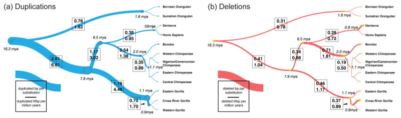
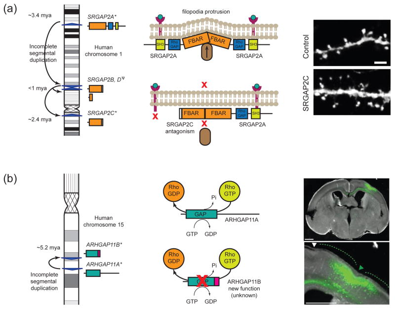
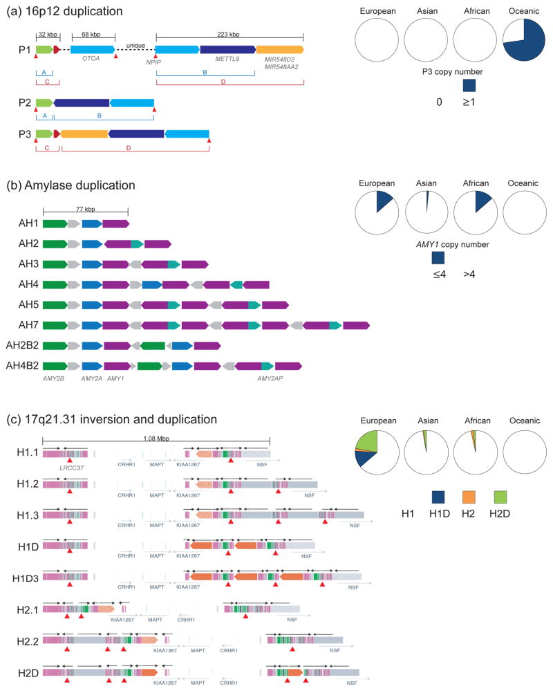
Duplications are the primary force by which new gene functions arise and provide a substrate for large-scale structural variation. Analysis of thousands of genomes shows that humans and great apes have more genetic differences in content and structure over recent segmental duplications than any other euchromatic region. Novel human-specific duplicated genes, ARHGAP11B and SRGAP2C, have recently been described with a potential role in neocortical expansion and increased neuronal spine density. Large segmental duplications and the structural variants they promote are also frequently stratified between human populations with a subset being subjected to positive selection. The impact of recent duplications on human evolution and adaptation is only beginning to be realized as new technologies enhance their discovery and accurate genotyping.


Figures

Figure 1
Figure 1. Primate rates of duplication and deletion
Rates of fixed (a) duplication and (b) deletion are shown as a function of the number of substitutions along each branch of the great ape phylogeny. Branch widths are scaled proportionally to the number of duplicated base pairs per substituted base pair based on analysis of 97 human/ape genomes. A burst of duplicated base pairs appears to have occurred in the common ancestral branch leading to humans and African great apes, where duplicated base pairs were added at 2.6-fold the rate of substitution. In contrast, the rate of deletion in the great ape lineage is more clocklike along all branches (mean of 0.32 deleted base pairs per substitution) with the exception of the chimpanzee–bonobo ancestral lineage, where an approximate twofold increase in the rate of deletion is observed (0.71 deleted base pairs per substitution). Adapted from [10].
Figure 2
Figure 2. Gene duplication and neuroanatomical adaptations
(a) SRGAP2A encodes a 1,071 aa protein with three protein domains shown as boxes: FBAR (orange), RhoGAP (blue), and SH3 (green). An incomplete SD from chromosome 1q32.1 created SRGAP2B at 1q21.1 encoding a partial FBAR domain (458 aa) and seven unique residues [33]. A subsequent duplication from SRGAP2B created SRGAP2C at 1p12.1 and SRGAP2D at 1q21.1 (which later partially deleted and represents a pseudogene). SRGAP2A homodimerizes and assembles at the cell membrane surface via its SH3 domain and induces filopodia protrusion by interacting with a protein complex including F-actin (brown oval) [37]. Truncated SRGAP2C is capable of heterodimerizing with SRGAP2A via its FBAR domain but lacks the RhoGAP and SH3 domains in turn antagonizing the function of the ancestral gene by not allowing it to assemble at the cell membrane surface. SRGAP2C expression induces long thin spines in mouse-cultured cortical neurons that phenocopies SRGAP2A deficiency in mice. Pictured are segments of dendrites from cortical neurons (20DIV) expressing EGFP alone (control) or EGFP and SRGAP2C (SRGAP2C) imaged two days after transfection. Reprinted with permission from [32]. (b) ARGHAP11A encodes a 1023 aa protein with a RhoGAP domain (blue). It was partially duplicated at chromosome 15q13.3 resulting in a paralog ARHGAP11B encoding a truncated RhoGAP domain (220 aa) and 47 unique residues at the C terminus (pink box) [35]. Via its RhoGAP domain, ARHGAP11A and truncated alternative isoforms encoding 220 aa (not pictured) dephosphorylate Rho-GTP unlike ARHGAP11B, which does not exhibit RhoGAP activity. ARHGAP11B overexpression leads to an increase in basal progenitors in the mouse neocortex possibly inducing cortical folding. Pictured are coronal sections of an E18.5 mouse telencephali in utero electroporated at E13.5 with ARHGAP11B and GFP expression plasmids. Phase contrast and GFP fluorescence of one section along the rostro-caudal axis. Scale bars, 500 μm. Green and white dashed lines and triangles indicate gyrus- and sulcus-like structures in and adjacent to the electroporated area, respectively. Reprinted with permission from [34].
Figure 3
Figure 3. Population diversity of duplications in modern human populations
SDs are indicated as colored blocks with directionality shown. Pie charts show frequencies of paralogs/alleles across European (E), Asian (As), African (Af), and Oceanic (O) populations. Core duplicons (red triangles) are depicted below structural paralogs or haplotypes. (a) A ~255 kbp Oceanic-specific SD (P3) was identified and shown to be made up of disperse segments from chromosome 16p12.1. The duplication is present in the genome of the ancient hominin, Denisova, but not observed in any other modern human populations possibly as a result of introgression back into the human lineage. The Oceanic-specific P3 formed as a result of interspersed duplications of P1 (represented in the human reference genome GRCh38) and P2 and included the NPIP core duplicon. P3 frequency was estimated based on genome sequence read-depth from the Human Genome Diversity Project (HGDP) cohort (E = 59, As = 45, Af = 36, O = 21) [41]. The P3 duplication has been identified in all Papuan individuals. (b) Diverse duplication structures exist at the amylase locus, with at least eight predicted haplotypes representing varying copies of AMY2B, AMY2A, AMY1, and AMY2AP (figure adapted from [50]). Overall copy number estimates of AMY1 in 1000 Genomes Project [41] and HGDP cohorts [44] were calculated and low-copy (CN ≤ 4) versus high-copy (CN > 4) frequencies were determined (E = 145, As = 204, Af = 133, O = 21). European and African populations show overall lower copy numbers of AMY1 compared with Asian and Oceanic. (c) Structural diversity of the chromosome 17q21.31 haplotype (figure adapted from [54]). Various forms of the directly orientated haplotype (H1) have been identified, including European-enriched haplotypes that show duplications of the promoter and first exon of KANSL1 (H1D). The inverted haplotype (H2) exists in a simpler form (H2.1) found among the San Khoisan and in more complex duplicated forms in European/Mediterranean haplotypes, including a smaller duplication of the promoter and first exon of KANSL1 (H2D). Allele frequencies of H1, H1D, H2, and H2D are shown based on sequence data from 1000 Genomes Project and HGDP cohorts (E = 628, As = 733, Af = 820, O = 27) [54]. H2.1 is predicted to represent the ancestral haplotype (2.3 mya), H1 is now the dominant haplotype worldwide, and the increased frequencies of H1D, H2, and H2D in European populations are the result of positive selection or extraordinary genetic drift. This is remarkable in light of the fact that the European H2 haplotype is predisposed to microdeletion due to the accumulation of directly oriented SDs. The less complex pattern of SDs observed among African H2.1 allele carriers suggest that the ancestral H2 haplotype is not predisposed to disease [54].

References

    1. Bailey JA, Yavor AM, Massa HF, Trask BJ, Eichler EE. Segmental duplications: organization and impact within the current human genome project assembly. Genome Res. 2001;11:1005–1017. - PMC - PubMed
    1. Haldane JBS. The causes of evolution. London, New York etc: Longmans, Green and co; 1932.
    1. Ohno S. Evolution by gene duplication. London, New York: Allen & Unwin; Springer-Verlag; 1970.
    1. Lynch M, Conery JS. The evolutionary fate and consequences of duplicate genes. Science. 2000;290:1151–1155. - PubMed
    1. Carvalho CM, Lupski JR. Mechanisms underlying structural variant formation in genomic disorders. Nat Rev Genet. 2016;17:224–238. This review provides an excellent resource of currently known mechanisms of structural variant formation leading to SDs. - PMC - PubMed