Review
. 2016 Oct 26;116(20):12688-12710.
doi: 10.1021/acs.chemrev.6b00296. Epub 2016 Sep 1.

Functions and Malfunctions of Mammalian DNA-Cytosine Deaminases

Affiliations
Review

Functions and Malfunctions of Mammalian DNA-Cytosine Deaminases

Sachini U Siriwardena et al. Chem Rev. .

Abstract

The AID/APOBEC family enzymes convert cytosines in single-stranded DNA to uracils, causing base substitutions and strand breaks. They are induced by cytokines produced during the body's inflammatory response to infections, and they help combat infections through diverse mechanisms. AID is essential for the maturation of antibodies and causes mutations and deletions in antibody genes through somatic hypermutation (SHM) and class-switch recombination (CSR) processes. One member of the APOBEC family, APOBEC1, edits mRNA for a protein involved in lipid transport. Members of the APOBEC3 subfamily in humans (APOBEC3A, APOBEC3B, APOBEC3C, APOBEC3D, APOBEC3F, APOBEC3G, and APOBEC3H) inhibit infections of viruses such as HIV-1, HBV, and HCV, and retrotransposition of endogenous retroelements through mutagenic and nonmutagenic mechanisms. There is emerging consensus that these enzymes can cause mutations in the cellular genome at replication forks or within transcription bubbles depending on the physiological state of the cell and the phase of the cell cycle during which they are expressed. We describe here the state of knowledge about the structures of these enzymes, regulation of their expression, and both the advantageous and deleterious consequences of their expression, including carcinogenesis. We highlight similarities among them and present a holistic view of their regulation and function.


Figures
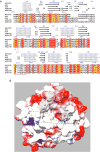
Figure 1
Figure 1
Structure of AID/APOBEC family proteins. A. Multiple sequence alignment of human APOBEC3 proteins with known crystal structures. ENDscript 2 software was used to generate the sequence alignment of A3A (PDB: 4XX0), A3B-CTD (PDB: 5CQD), A3C (PDB: 3VM8) and A3G-CTD (PDB: 3IR2) using A3F-CTD (PDB: 4IOU) as the query. It is important to note that some of the crystallized proteins had several amino acid substitutions and in cases of proteins with two Zn2+ binding domains, only the carboxy-terminal domain (CTD) was crystallized. Residues shown in white against a red background are identical and similar residues are shown in red against a yellow background. The amino acid residues that constitute the active site are marked with red asterisks and the putative DNA binding region is identified with a dashed red line. Secondary structure elements in all APOBEC3 crystal structures are displayed above the sequence alignment (α helices with squiggles, β strands with arrows and turns with the letter T). Helices are numbered from α1 through α6, strands from β1 through β5 and loop regions from L1 through L10. B. The Coulombic surface potential of the A3F-CTD structure was generated using UCSF Chimera software. The color code is blue (positive), white (neutral) and red (negative). C280, C283 and H249 residues coordinate Zn ion (magenta). Active site glutamate (E251) is also shown. L7 (yellow) is the putative DNA sequence recognition loop and two possible trajectories for DNA binding are shown as dashed green lines.
Figure 2
Figure 2
Role of AID in antibody maturation. Schematic organization of the murine heavy chain gene (IgH) and its alterations during antibody maturation are shown. Abbreviations- VDJ- Rearranged V, D and J segments; E, enhancer, S, switch region; C, constant region; 3′-RR, regulatory region, Pol II, RNA polymerase II; AID, activationinduced deaminase; UNG2, nuclear form of UNG; AP endonuclease, abasic endonuclease; MMR, mismatch repair; TLS polymerase, translesion synthesis polymerase; NHEJ, non-homologous end-joining; altEJ, alternate end-joining. The subscripts μ, ε and α to different constant segments. Representative base substitution mutations within VDJ (SHM) and the μ-ε hybrid switch region are shown and the IgM to IgE isotype switch in the antibody is indicated in the figure.
Figure 3
Figure 3
Consequences of cytosine deamination. A. Consequences of cytosine deamination in a transcription bubble. The direction of transcription is shown by an arrow and the mRNA is shown in green. During transcription the cytosines in the non-template strand of the transcription bubble are accessible and may be deaminated by the AID/APOBEC proteins. The resulting uracils may be repaired through error-free base excision repair restoring the C:G pair, copied during replication creating G:C to A:T mutations or undergo error-prone repair resulting in all possible base substitutions. N: any base, N′: complement of N. B. Consequences of cytosine deamination at a replication fork. A replication fork with cytosines in the leading strand template (LDST) and the lagging strand template (LGST) are shown. The closed arrow-head represents the helicase that opens the DNA and is pointed in the overall direction of replication. AID/APOBEC proteins deaminate cytosines in LGST and the resulting uracils are either copied during replication (upper branch) or undergo repair (lower branch). Replication without repair results in C:G to T:A mutations in the LGST. Alternately, uracil-DNA glycosylase (UNG) may excise the uracils and the resulting abasic sites may be copied by translesion synthesis (TLS) polymerases creating mutations (downward arrow). The predominant mutations caused by polymerases η and REV1 are shown. AP endonuclease may nick the DNA at the abasic site and this will lead to double strand (DS) breaks in DNA and cause replication fork to collapse.
Figure 4
Figure 4
Linking viral infection, AID/APOBEC expression and cancer. During viral infection cytokines (yellow triangles) trigger the expression of AID/APOBEC proteins (purple ovals) via NF- κB pathway. These enzymes damage the viral genome (red star) and promote creation of high affinity antibodies against the virus. This helps the body clear the virus. Additionally, off-target damage to cellular DNA leads to mutations and strand breaks resulting in genome instability. This may lead to cell death or malignant transformation.

References

    1. Beale RC, Petersen-Mahrt SK, Watt IN, Harris RS, Rada C, Neuberger MS. Comparison of the differential context-dependence of DNA deamination by APOBEC enzymes: correlation with mutation spectra in vivo. Journal of molecular biology. 2004;337:585. - PubMed
    1. Bransteitter R, Pham P, Scharff MD, Goodman MF. Activation-induced cytidine deaminase deaminates deoxycytidine on single-stranded DNA but requires the action of RNase. Proceedings of the National Academy of Sciences of the United States of America. 2003;100:4102. - PMC - PubMed
    1. Chaudhuri J, Tian M, Khuong C, Chua K, Pinaud E, Alt FW. Transcription-targeted DNA deamination by the AID antibody diversification enzyme. Nature. 2003;422:726. - PubMed
    1. Dickerson SK, Market E, Besmer E, Papavasiliou FN. AID mediates hypermutation by deaminating single stranded DNA. The Journal of experimental medicine. 2003;197:1291. - PMC - PubMed
    1. Petersen-Mahrt SK, Neuberger MS. In vitro deamination of cytosine to uracil in single-stranded DNA by apolipoprotein B editing complex catalytic subunit 1 (APOBEC1) The Journal of biological chemistry. 2003;278:19583. - PubMed