

Comment
doi: 10.1038/cr.2016.104.
Epub 2016 Sep 2.
Presenting mitochondrial antigens: PINK1, Parkin and MDVs steal the show
Affiliations
- PMID: 27585536
- PMCID: PMC5099861
- DOI: 10.1038/cr.2016.104
Item in Clipboard
Comment
Presenting mitochondrial antigens: PINK1, Parkin and MDVs steal the show
Cell Res.
2016 Nov.
Abstract
In a recent paper published in Cell, Matheoud et al. demonstrated that, in response to cellular stress, self-antigens can be extracted from mitochondria via mitochondrial-derived vesicles and presented at the cell surface to trigger an immune response; this pathway, termed mitochondrial antigen presentation (MitAP), is repressed by PINK1 and Parkin. These findings implicate autoimmune mechanisms in Parkinson's disease.
Figures
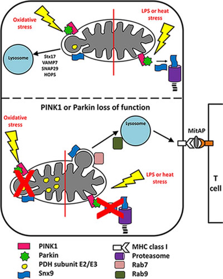
The role of PINK1, Parkin and MDVs in MitAP. (Top panel, right) Under conditions of heat stress or treatment with LPS, PINK1 and Parkin repress MitAP by mediating the proteasomal turnover of Snx9. (Top panel, left) Oxidative stress induces the generation of PDH-positive MDVs containing oxidized and damaged cargo that are directed to lysosomes for degradation. PINK1, Parkin and Snx9 are required for their formation, and Stx17, VAMP7, SNAP29 and the HOPS complex are required for their fusion with the lysosome. (Bottom panel, left) In cells lacking functional PINK1 or Parkin, oxidative stress fails to induce the formation of PDH-positive MDVs. (Bottom panel, right) In contrast, treatment with LPS or heat stress in PINK1- or Parkin- null cells induces the formation of MDVs harboring mitochondrial antigens, which requires Snx9 and Rab7 for their formation and Rab9 for fusion with the lysosome. Antigens are processed in the lysosome for presentation on MHC class I molecules at the cell surface. MitAP cognate CD8+ T cells are activated upon binding of their T cell receptors to MHC class I molecules presenting mitochondrial antigens.
Comment on
-
Parkinson's Disease-Related Proteins PINK1 and Parkin Repress Mitochondrial Antigen Presentation.Cell. 2016 Jul 14;166(2):314-327. doi: 10.1016/j.cell.2016.05.039. Epub 2016 Jun 23. Cell. 2016. PMID: 27345367
References
-
- Matheoud D, Sugiura A, Bellemare-Pelletier A, et al. Cell 2016; 166:314–327. - PubMed
Publication types
MeSH terms
Substances
LinkOut - more resources
Full Text Sources
Other Literature Sources
Medical
