. 2016 Sep 1;13(1):226.
doi: 10.1186/s12974-016-0689-1.

Cellular investigations with human antibodies associated with the anti-IgLON5 syndrome

Affiliations

Cellular investigations with human antibodies associated with the anti-IgLON5 syndrome

Lidia Sabater et al. J Neuroinflammation. .

Abstract

Background: Antibodies against IgLON5, a neuronal adhesion protein of unknown function, are markers of a novel neurological disorder termed anti-IgLON5 syndrome. The disorder shows a remarkable association with the HLA-DQB1*0501 and HLA-DRB1*1001 alleles, and postmortem studies demonstrate a novel neuronal tauopathy predominantly involving the hypothalamus and tegmentum of the brainstem. The role of IgLON5 antibodies in the pathogenesis of the disease is currently unknown. Here, we have determined the target epitopes of IgLON5 antibodies, the effects of the IgLON5 antibodies in rat hippocampal neurons, and the IgG subclass responsible for these effects.

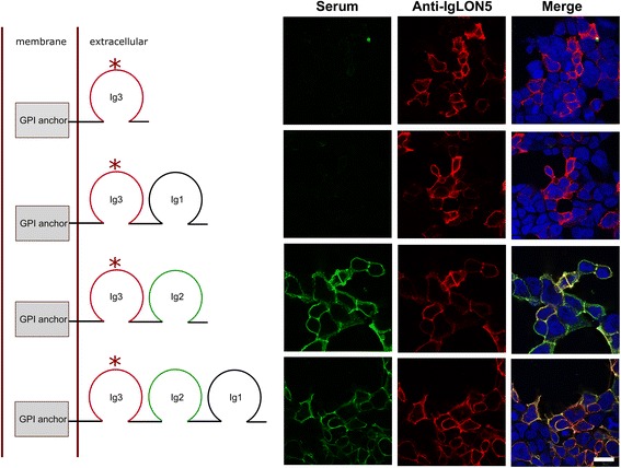
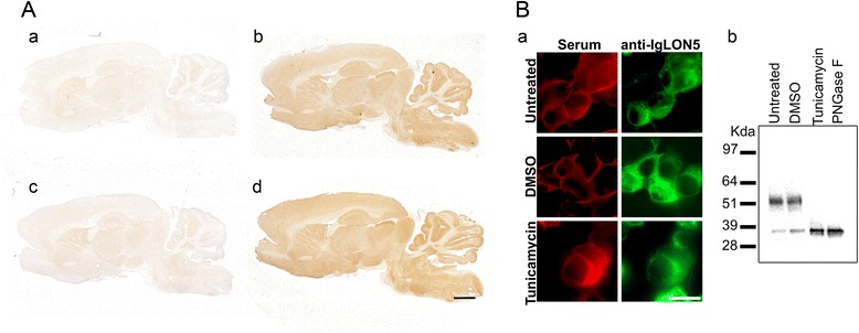
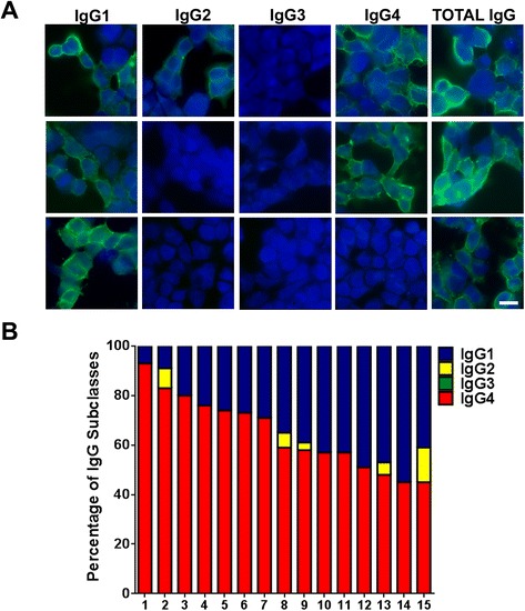
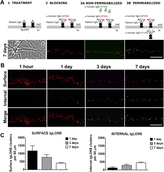
Methods: HEK293 cells expressing several deletion constructs of IgLON5 were used to determine the epitopes recognized by the serum of 15 patients with anti-IgLON5 syndrome. The role of glycosylation in immunogenicity was tested with PNGase F treatment of transfected cells. Dissociated hippocampal neuronal cultures were used to test by immunocytochemistry the effects of total IgG, IgG1, and IgG4 subclasses of IgLON5 antibodies.

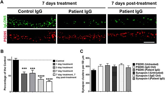
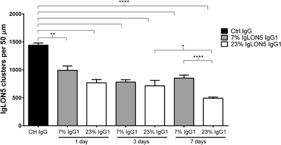
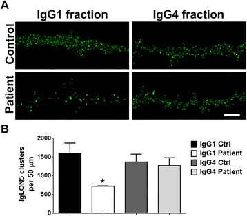
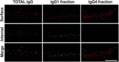
Results: Patients' antibodies reacted with the immunoglobulin-like domain 2 of IgLON5. Glycosylation was not required for immunoreactivity. The predominant subclass of IgLON5 antibodies was IgG4 but all patients also had IgG1. The mean percentage of specific IgLON5 IgG4 and IgG1 of the samples analyzed by flow cytometry was 64 and 33 %, respectively. In cultures of hippocampal neurons, patients' antibodies caused a decrease of cell surface IgLON5 clusters that was not reversed after IgLON5 antibodies were removed from the media. The decrease of surface IgLON5 clusters correlated with the rate of antibody internalization. These effects were observed with purified IgG1 but not with the IgG4 antibodies.

Conclusions: IgLON5 antibodies recognize the immunoglobulin-like domain 2 of the antigen, and the reactivity is not dependent on glycosylation. The effects observed on hippocampal neuronal cultures indicate an irreversible antibody-mediated internalization of surface IgLON5. These effects were mediated by specific IgLON5 IgG1 antibodies and suggest a pathogenic role of these antibodies in the disease.

Keywords: Autoantibodies; IgLON5; Immunoglobulin subclasses; Tauopathy.


Figures

Fig. 1
Fig. 1
Epitope analysis of IgLON5 antibodies. The diagrams depict the complete IgLON5 (bottom) and mutated clones (left). Immunofluorescence of transfected HEK293 cells with the full length or the mutant IgLON5 clones (right). Rows correspond to the indicated clone in the diagram. The serum with IgLON5 antibodies (green) only targets the clones containing the immunoglobulin-like domain 2 (Ig2, left column). IgLON5 commercial antibody immunoreactivity is shown in the central column (in red) and both reactivities are shown merged in the right column. Nuclei counterstained with DAPI (blue). Scale bar = 10 μm. The (*) indicates the epitope recognized by the IgLON5 commercial antibody
Fig. 2
Fig. 2
Deglycosylation assay. Western blot of human cerebellum protein extract treated with N-Gly (N-glycosidase), O-Gly (O-glycosidase), and/or NA (neuraminidase) and probed with a commercial rabbit anti-human IgLON5 antibody. The IgLON5 protein is completely deglycosylated only when the N-linked glycans are removed reaching its predicted molecular weight of 36 kDa according to the amino acid sequence
Fig. 3
Fig. 3
Deglycosylated IgLON5 is recognized by human IgLON5 antibodies. A Anti-IgLON5 immunoreactivity in rat brain sections is totally abrogated when the anti-IgLON5-positive serum is preabsorbed with glycosylated (a) or deglycosylated protein extracts from transfected HEK293 with IgLON5 (c). The immunoreactivity of IgLON5 antibodies is present when the serum is preabsorbed with an unrelated antigen glycosylated (b) or deglycosylated (d). Scale bar = 1000 μm. B a Immunofluorescence of HEK293 cells transfected with IgLON5. Prevention of N-glycosylation with tunicamycin treatment does not affect the reactivity of patients’ antibodies with IgLON5. Scale bar = 10 μm. b Western blot of extracts of the indicated IgLON5-HEK293 cells shown in (a), demonstrating a shift of the molecular weight (from ~52 to 36 kDa) consistent with the deglycosylation of the protein. The findings confirm that the reactivity of patients’ serum antibodies with tunicamycin-treated cells of A was directed against deglycosylated epitopes
Fig. 4
Fig. 4
Analysis of IgLON5 antibody subclasses. a Example of anti-IgLON5 IgG subclasses in three different sera. HEK293 cells were transfected with IgLON5 and incubated with three sera with IgLON5 antibodies followed by antibodies against total IgG or specific IgG subclasses. Positive immunoreactivity is shown in green. Nuclei counterstained with DAPI (blue). Scale bar = 10 μm. b IgG subclass percentages of IgLON5 antibodies in the 15 positive serum samples analyzed by flow cytometry
Fig. 5
Fig. 5
IgLON5 antibodies irreversible decrease IgLON5 clusters on cell surface. a Immunofluorescence on hippocampal neurons treated for 7 days (14DIV to 21DIV) with control or patient with anti-IgLON5 IgG. The surface clusters are drastically reduced by the anti-IgLON5 IgG, and the effect is irreversible because 7 days after removing the anti-IgLON5 IgG the reduced number of surface IgLON5 clusters persisted. Scale bar = 5 μm. b Quantification of the decrease of IgLON5 clusters on the dendrite surface in a time-course treatment of 1, 3, and 7 days, and 7 days after removing the anti-IgLON5 IgG. ***p < 0.005, ****p < 0.0001. c Synaptic markers (PSD95, synapsin-I) were not affected by the anti-IgLON5 IgG treatment
Fig. 6
Fig. 6
Comparison of the time-dependent decrease of neuronal cell surface IgLON5 clusters after treatment with total IgG of two patients with different levels of anti-IgLON5 IgG1 antibodies. On days 1 and 3, treatment with IgG of the patient with the lower level of IgG1 IgLON5 antibodies (7 % of total IgLON5 antibodies) produced a similar decrease of IgLON5 clusters as the IgG of the patient with average level of IgG1 antibodies (23 % of total IgLON5 antibodies). (**p = 0.001, ****p < 0.0001, comparing with control IgG, one-way ANOVA with Bonferroni’s multiple comparison test). In contrast, on day 7, the sample of the patient with lower levels of IgG1 antibodies did not decrease further the levels of IgLON5, whereas the sample of the patient with average level of IgG1 IgLON5 antibodies produced an additional decrease of IgLON5 clusters (****p < 0.0001) that was statistically significant comparing with day 3 (*p = 0.03)
Fig. 7
Fig. 7
IgLON5 IgG1, but not IgG4 antibodies, cause a reduction of surface IgLON5 clusters. a Hippocampal neurons were treated for 3 days with IgG1 or IgG4 fractions from control serum or from a patient with IgLON5 antibodies and immunostained for surface antibody-bound IgLON5. Scale bar = 5 μm. b Quantification of IgLON5 clusters. IgLON5 clusters were significantly reduced after treatment with patient IgG1 compared with control IgG1 or patient and control IgG4. *p < 0.05
Fig. 8
Fig. 8
IgLON5 antibodies produce internalization of IgLON5 clusters. a Panel 1: Hippocampal neurons treated for 7 days with IgG-positive IgLON5 antibodies. Panel 2: IgG bound to IgLON5 on the dendrite surface labeled live with an excess of anti-human IgG Alexa Fluor 594 (red). Panel 3A: The saturation of the neuronal surface prevents that the anti-human IgG Alexa Fluor 488 (green) attaches to the surface. Panel 3B: After the neuron is permeabilized and incubated with the anti-human IgG Alexa Fluor 488, the green fluorescence localizes the human IgG attached to internalized IgLON5 clusters. Scale bar = 5 μm. b The internalization of IgLON5 clusters is a time-dependent effect and parallels a decrease of IgLON5 clusters on the surface of the dendrite. c Quantification of the number of IgLON5 clusters remaining on the cell surface and internalized after the treatment
Fig. 9
Fig. 9
IgG1 IgLON5 antibodies internalize IgLON5 clusters. Hippocampal neurons treated for 3 days with total IgG, IgG1, and IgG4 IgLON5 antibodies. The immunofluorescence strategy to differentiate surface and internal human IgG bound to IgLON5 is conducted as in Fig. 8. The IgG1 antibodies alone could reproduce the same effects seen with the total IgG; meanwhile, the IgG4 did not produce internalization. Scale bar = 5 μm

References

    1. Sabater L, Gaig C, Gelpi E, Bataller L, Lewerenz J, Torres-Vega E, et al. A novel non-rapid-eye movement and rapid-eye-movement parasomnia with sleep breathing disorder associated with antibodies to IgLON5: a case series, characterisation of the antigen, and post-mortem study. Lancet Neurol. 2014;13:575–86. doi: 10.1016/S1474-4422(14)70051-1. - DOI - PMC - PubMed
    1. Simabukuro MM, Sabater L, Adoni T, Cury RG, Haddad MS, Moreira CH, et al. Sleep disorder, chorea, and dementia associated with IgLON5 antibodies. Neurol Neuroimmunol Neuroinflamm. 2015;2:e136. doi: 10.1212/NXI.0000000000000136. - DOI - PMC - PubMed
    1. Högl B, Heidbreder A, Santamaria J, Graus F, Poewe W. IgLON5 autoimmunity and abnormal behaviours during sleep. Lancet. 2015;385:1590. doi: 10.1016/S0140-6736(15)60445-7. - DOI - PubMed
    1. Brüggemann N, Wandinger KP, Gaig C, Sprenger A, Junghanns K, Helmchen C, et al. Dystonia, lower limb stiffness, and upward gaze palsy in a patient with IgLON5 antibodies. Mov Disord. 2016 - PubMed
    1. Eisenhaber B, Bork P, Eisenhaber F. Sequence properties of GPI-anchored proteins near the omega-site: constraints for the polypeptide binding site of the putative transamidase. Protein Eng. 1998;11:1155–61. doi: 10.1093/protein/11.12.1155. - DOI - PubMed

Publication types

MeSH terms