. 2016 Sep 2:7:12706.
doi: 10.1038/ncomms12706.

Nestin(+) cells direct inflammatory cell migration in atherosclerosis

Affiliations

Nestin(+) cells direct inflammatory cell migration in atherosclerosis

Raquel Del Toro et al. Nat Commun. .

Abstract

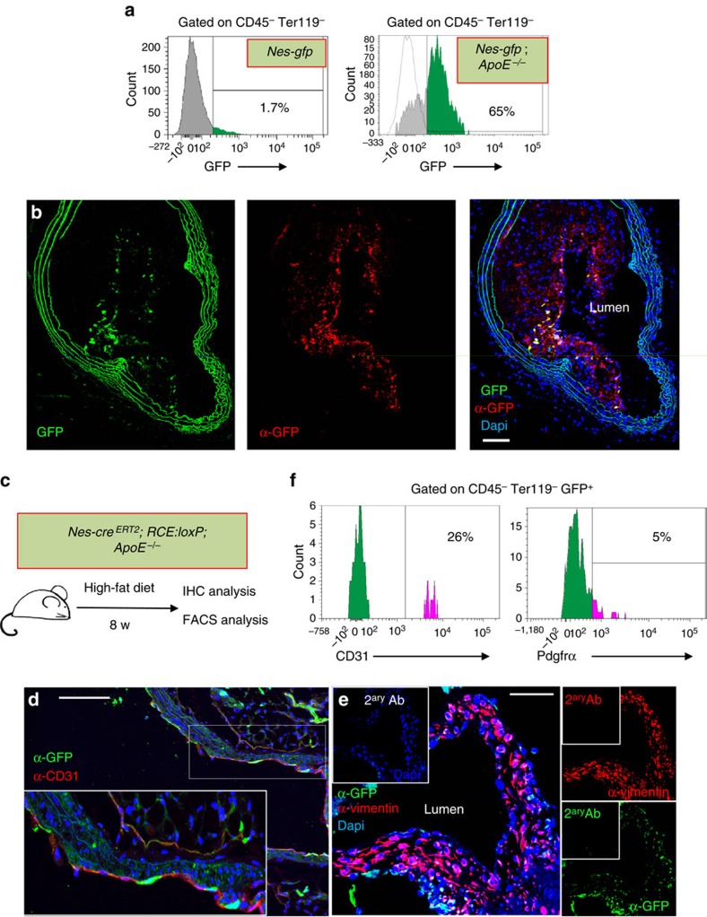
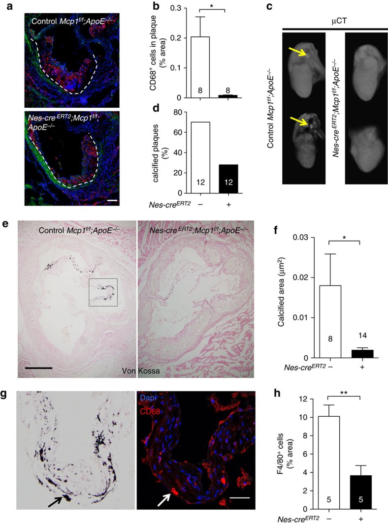
Atherosclerosis is a leading death cause. Endothelial and smooth muscle cells participate in atherogenesis, but it is unclear whether other mesenchymal cells contribute to this process. Bone marrow (BM) nestin(+) cells cooperate with endothelial cells in directing monocyte egress to bloodstream in response to infections. However, it remains unknown whether nestin(+) cells regulate inflammatory cells in chronic inflammatory diseases, such as atherosclerosis. Here, we show that nestin(+) cells direct inflammatory cell migration during chronic inflammation. In Apolipoprotein E (ApoE) knockout mice fed with high-fat diet, BM nestin(+) cells regulate the egress of inflammatory monocytes and neutrophils. In the aorta, nestin(+) stromal cells increase ∼30 times and contribute to the atheroma plaque. Mcp1 deletion in nestin(+) cells-but not in endothelial cells only- increases circulating inflammatory cells, but decreases their aortic infiltration, delaying atheroma plaque formation and aortic valve calcification. Therefore, nestin expression marks cells that regulate inflammatory cell migration during atherosclerosis.


Figures

Figure 1
Figure 1. Nestin+ cells regulate inflammatory cell traffic in atherosclerosis.
(a) Flow cytometry plots showing the cell sorting strategy. (b) QPCR analysis of Icam1, Vcam1 and Mcp1 mRNA in BM CD45 Ter119 CD31+ endothelial cells (BMEC) and BM CD45 Ter119 CD31 stromal cells (BMSC) from ApoE−/− mice fed with chow or HFD for 2 months (n=3–7). (c) CD31 expression in BM GFP+ stromal cells from Nes-gfp;ApoE−/− mice fed with HFD for 2–3 months (n=4). (d) Experimental design of peripheral blood analysis from mice treated with tamoxifen and fed with HFD for 2 months. (e,f) Flow cytometry strategy used to identify inflammatory neutrophils, inflammatory monocytes and non-classical monocytes. (g,h) Number of non-classical monocytes, inflammatory monocytes and inflammatory neutrophils in the peripheral blood of tamoxifen-treated (g) Nes-CreERT2;Mcp1f/f;ApoE−/− mice, (h) Cdh5-CreERT2;Mcp1f/f;ApoE−/− mice and control littermates (n=3–12). Data are means±s.e.m.; n and P values are indicated; *P<0.05, unpaired two-tailed t test.
Figure 2
Figure 2. Mcp1 deletion in nestin+ cells delays atherosclerosis progression.
(a,c) Representative photographs of whole-mounted aortas from tamoxifen-treated (a) Cdh5-creERT2;Mcp1f/f;ApoE−/− mice, (c) Nes-creERT2;Mcp1f/f;ApoE−/− mice and control littermates (without Cre) two months after HFD, stained with Oil Red O (red) to mark atheroma plaques (n=6). (a,c) Scale bar, 1 mm. (b,d) Lesion size and coverage in the aortic arch (left) and number of lesions in the thoracic aorta (right) of these mice (n=3–6). Data are means±s.e.m.; n and P values are indicated; *P<0.05, unpaired two-tailed t test.
Figure 3
Figure 3. Mcp1 deletion in nestin+ cells decreases the inflammatory infiltration in the aortic wall.
(ad) QPCR analysis of Mcp1, Cxcl12 and Icam1 mRNA in aorta from mice fed with chow after the tamoxifen injection (n=6–12). (e) Flow cytometry plots of aortic cells (left) and myeloid cells (right). (f) Representative flow cytometry diagrams of the aortas depicting inflammatory neutrophils (CD11b+ Ly6Ghigh, red) and inflammatory monocytes (CD11b+ Ly6Chigh, green) in both groups of mice. Frequency of (g) hematopoietic cells (n=9–12), (h) inflammatory neutrophils (n=10–13) and (i) inflammatory monocytes in the aortas of Nes-creERT2;Mcp1f/f;ApoE−/− mice and control littermates fed with HFD for 2 months (n=9–12). (j) Design of adoptive transfer experiments. BM Gr1+ leukocytes and peripheral blood mononuclear cells (PBMCs) from CD45.1 mice fed with HFD were intravenous transplanted into Nes-creERT2;Mcp1f/f;ApoE−/− mice and control littermates previously treated with tamoxifen and fed with HFD for 2 months. (k) Representative flow cytometry diagrams of CD45.1+ and CD45.2+ cells in the aortas of Nes-creERT2;Mcp1f/f;ApoE−/− mice and control littermates. (l) Frequency of CD45.1+ cells in the BM, the peripheral blood (PB) and the aorta of both groups of mice (n=6–15). (e,f,k) The frequencies of the gated populations are indicated. Data are means±s.e.m.; n and P values are indicated; *P<0.05, unpaired two-tailed t test.
Figure 4
Figure 4. Nestin+ cells in the aortic adventitia and media express Mcp1 in mice and humans.
Immunofluorescence of sections from (a,b,f) thoracic aorta and (c) aortic valves from Nes-Gfp mice using antibodies against (a,b) GFP and (c) CD31 (red). Note that nestin+ cells are mainly distributed in the adventitia (b) or in close proximity of valve endothelial cells (c). Inset, high magnification of the aortic leaflets. (d,e) Immunofluorescence of representative control section from WT thoracic aorta using antibodies against GFP (red). The absence of red signal demonstrates the specificity of the staining. (b,e) The autofluorescence of the smooth muscle layer is shown in green. (f) Negative control immunofluorescence on Nes-gfp aorta using control IgG (top panels) and/or secondary antibodies (bottom panels). (b,c,e,f) Nuclei were counterstained with DAPI (blue). (gj) Immunofluorescence of (gi) NESTIN and (hj) MCP1 in consecutive cross sections from human (g,h) coronary and (i,j) carotid artery samples. Red arrowheads depict cells that express both proteins in consecutive sections. (a,b,df,gj) Scale bars, 50 μm, (c) 100 μm.
Figure 5
Figure 5. Nestin+ cells participate in the formation of the atheroma plaque.
(a) Flow cytometry of aortic stromal cells showing GFP expression in Nes-gfp mice fed with chow diet (left panel) and in Nes-gfp;ApoE−/− mice fed with HFD for 2 months (right panel). (b) Representative GFP immunofluorescence of a section of the brachiocephalic artery branching out of the aortic arch of Nes-Gfp;ApoE−/− mice fed with HFD for 2 months. Abundant GFP+ cells were found in the atheroma plaque and the adventitial layer. GFP was detected with an anti-GFP antibody (red). Nuclei were counterstained with DAPI (blue). (c) Experimental design of lineage-tracing studies. Nes-creERT2;Rosa26-Gfp;ApoE−/− mice were injected with tamoxifen to trace the progeny of nestin+ cells and were fed with HFD for 2 months. (d) Immunofluorescence of the aortic valves using anti-GFP and anti-CD31 antibodies. Nuclei were counterstained with DAPI (blue). Inset, higher magnification of aortic leaflet. (e) Immunofluorescence of the aortic valves with anti-vimentin antibody. Nuclei were counterstained with DAPI (blue). (f) Flow cytometry histogram showing Pdgfrα and CD31 expression in aortic CD45 Ter119 GFP+ cells. The frequencies of depicted populations are indicated. (be) Scale bar, 100 μm.
Figure 6
Figure 6. Mcp1 deletion in nestin+ cells reduces vascular calcification.
(a) Immunofluorescence of CD68 (red) to label macrophages infiltrated in the valves of Nes-creERT2;Mcp1f/f;ApoE−/− mice and Mcp1f/f;ApoE−/− controls fed with HFD for 6 weeks. Nuclei were counterstained with DAPI (blue). (b) Quantification of CD68+ cells in the atheroma plaque of these mice (n=8). (c) Representative ex-vivo microtomography (μCT) photographs of the hearts of Nes-creERT2;Mcp1f/f;ApoE−/− mice and Mcp1f/f;ApoE−/− controls. Arrows indicate calcified plaques. (d) Frequency of hearts that showed calcified plaques (n=12). (e) Representative sections of Von Kossa staining of calcium deposits (black) in the aortic valves of these mice. (f) Calcified area in three sections from the aortic valves of these mice (n=8–14). (g) Consecutive aortic valves sections stained with Von Kossa (black) or DAPI (blue) and anti-CD68 antibodies (red). Note the proximity of CD68+ macrophages to the calcified areas (arrow, example). (h) Number of F4/80+ macrophages (not shown) infiltrated in the aortic leaflets (n=5). (b,d,f,h) Data are means±s.e.m.; n and P values are indicated; *P<0.05, **P<0.01, unpaired two-tailed t test. (a, g) Scale bars, 100 μm, (e) 200 μm.

Similar articles

Cited by

References

    1. Cybulsky M. I. & Gimbrone M. A. Jr. Endothelial expression of a mononuclear leukocyte adhesion molecule during atherogenesis. Science 251, 788–791 (1991). - PubMed
    1. Muller W. A. Mechanisms of leukocyte transendothelial migration. Annu. Rev. Pathol. 6, 323–344 (2011). - PMC - PubMed
    1. Randolph G. J. Mechanisms that regulate macrophage burden in atherosclerosis. Circ. Res. 114, 1757–1771 (2014). - PMC - PubMed
    1. Piedrahita J. A., Zhang S. H., Hagaman J. R., Oliver P. M. & Maeda N. Generation of mice carrying a mutant apolipoprotein E gene inactivated by gene targeting in embryonic stem cells. Proc. Natl Acad. Sci. USA 89, 4471–4475 (1992). - PMC - PubMed
    1. Zhang S. H., Reddick R. L., Piedrahita J. A. & Maeda N. Spontaneous hypercholesterolemia and arterial lesions in mice lacking apolipoprotein E. Science 258, 468–471 (1992). - PubMed

Publication types