

Lymphatic imaging to assess rheumatoid flare: mechanistic insights and biomarker potential
- PMID: 27586634
- PMCID: PMC5009676
- DOI: 10.1186/s13075-016-1092-0
Lymphatic imaging to assess rheumatoid flare: mechanistic insights and biomarker potential
Abstract
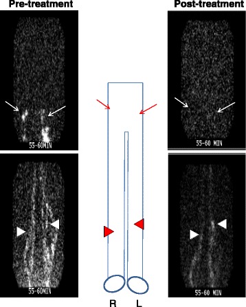
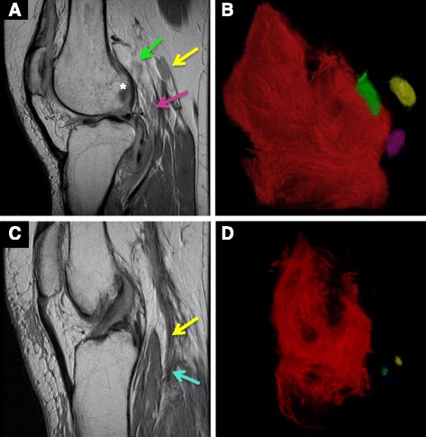
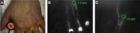
Proliferation of draining lymphatic vessels coupled with dynamic changes in lymph node volume and flow are characteristic features in rheumatoid arthritis (RA). Furthermore, impaired lymph egress from inflamed synovium is associated with joint flare in murine models of inflammatory-erosive arthritis. Unfortunately, advances towards a greater understanding of lymphatic changes in RA pathogenesis have been slow due to the absence of outcome measures to quantify lymphatic function in vivo. While lymphoscintigraphy is the current standard to assess lymphedema and sentinel lymph nodes in cancer patients, its sensitivity and specificity are inadequate to study lymphatics in RA. The emergence of high-resolution MRI, power Doppler ultrasound, and near-infrared imaging that permits real-time quantification of lymphatic function in animal models has been a major advance, and these techniques have produced a new paradigm of altered lymphatic function that underlies both acute arthritic flare and chronic inflammation. In acute flare, lymphatic drainage increases several fold, whereas no lymphatic contractions are detected in lymph vessels draining chronic arthritic joints. Moreover, these outcomes are now being adapted to study lymphatics in RA towards the development of novel biomarkers of arthritic flare and the discovery of new therapeutic targets. In particular, interventions that directly increase lymphatic egress from diseased joints by opening collateral lymphatic vessels, and that restore lymphatic vessel contractions, provide novel therapeutic approaches with potential for minimal toxicity and immunosuppression. To summarize the origins of this field, recent advances, and future directions, we herein review: current knowledge of lymphatics in RA based on classic literature; new in-vivo imaging modalities that have elucidated how lymphatics modulate acute versus chronic joint inflammation in murine models; and how these preclinical outcome measures are being translated to study lymphatic function in RA inflammation and how effective RA therapies alter lymphatic flow and lymph nodes draining flaring joints.
Trial registration: ClinicalTrials.gov NCT02680067 . Registered 7 December 2015; ClinicalTrials.gov NCT01098201 . Registered 30 March 2010; and ClinicalTrials.gov NCT01083563 . Registered 8 March 2010.
Figures
References
-
- Gibofsky A. Epidemiology, pathophysiology, and diagnosis of rheumatoid arthritis: a synopsis. Am J Manag Care. 2014;20(7):S128–35. - PubMed
-
- Carubbi F, Zugaro L, Cipriani P, Conchiglia A, Gregori L, Danniballe C, Pistoia ML, Liakouli V, Ruscitti P, Ciccia F, et al. Safety and efficacy of intra-articular anti-tumor necrosis factor alpha agents compared to corticosteroids in a treat-to-target strategy in patients with inflammatory arthritis and monoarthritis flare. Int J Immunopathol Pharmacol. 2016;29(2):252–66. doi: 10.1177/0394632015593220. - DOI - PMC - PubMed
-
- Lens JW, Vandenberg WB, Vandeputte LBA, Zwarts WA. Flare of antigen-induced arthritis in mice after intravenous challenge—kinetics of antigen in the circulation and localization of antigen in the arthritic and noninflamed joint. Arthritis Rheum. 1986;29(5):665–74. doi: 10.1002/art.1780290512. - DOI - PubMed
Publication types
MeSH terms
Associated data
Grants and funding
LinkOut - more resources
Full Text Sources
Other Literature Sources
Medical
