. 2016 Sep 2:6:32510.
doi: 10.1038/srep32510.

Replication Timing of Human Telomeres is Conserved during Immortalization and Influenced by Respective Subtelomeres

Affiliations

Replication Timing of Human Telomeres is Conserved during Immortalization and Influenced by Respective Subtelomeres

Laure Piqueret-Stephan et al. Sci Rep. .

Abstract

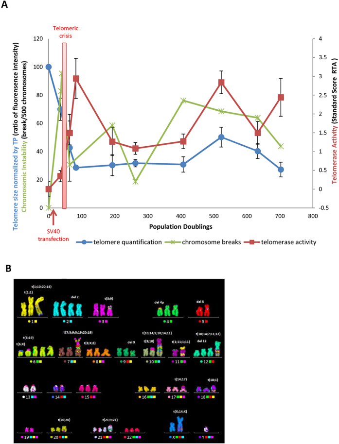
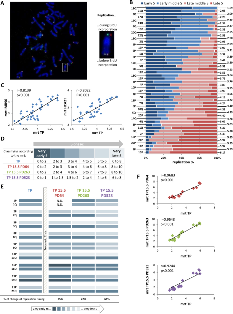
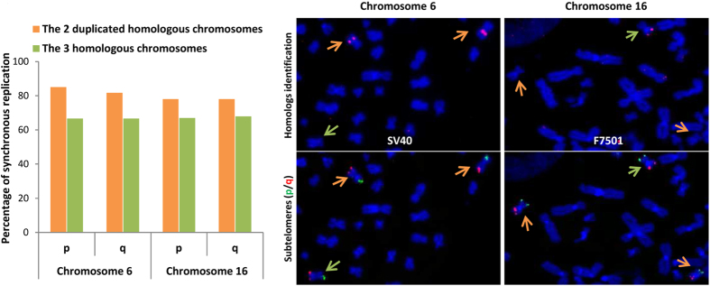
Telomeres are specific structures that protect chromosome ends and act as a biological clock, preventing normal cells from replicating indefinitely. Mammalian telomeres are replicated throughout S-phase in a predetermined order. However, the mechanism of this regulation is still unknown. We wished to investigate this phenomenon under physiological conditions in a changing environment, such as the immortalization process to better understand the mechanism for its control. We thus examined the timing of human telomere replication in normal and SV40 immortalized cells, which are cytogenetically very similar to cancer cells. We found that the timing of telomere replication was globally conserved under different conditions during the immortalization process. The timing of telomere replication was conserved despite changes in telomere length due to endogenous telomerase reactivation, in duplicated homologous chromosomes, and in rearranged chromosomes. Importantly, translocated telomeres, possessing their initial subtelomere, retained the replication timing of their homolog, independently of the proportion of the translocated arm, even when the remaining flanking DNA is restricted to its subtelomere, the closest chromosome-specific sequences (inferior to 500 kb). Our observations support the notion that subtelomere regions strongly influence the replication timing of the associated telomere.


Figures

Figure 1
Figure 1. Immortalization of TP fibroblasts following SV40 transfection.
(A) Evolution of mean telomere length (blue circles), the frequency of breaks per 500 chromosomes (green crosses) and telomerase activity (red squares) during cellular immortalization. (B) Example of chromosomal rearrangements in the TP 15.5 PD523 karyotype.
Figure 2
Figure 2. Variable length of S-phase and of its sub-phases during cellular immortalization.
(A) Example in TP cells of S-phase length determination with the aid of the observed distribution of the sub-phases at each BrdU incorporation time. The five S-sub-phases are in temporal order: Early S (E S), Early middle S (Em S), Middle S (M S), Late middle S (Lm S) and Late S (L S). (B) Reverse DAPI staining of metaphase spreads following ReD-FISH for the five S-sub-phases. (C) Summary of the length of each sub-phase for the control cells (TP) and for each population doubling studied (TP 15.5 PD64, PD263 and PD523). (D) To classify telomeres as a function of their replication, the mean replication timing (mrt) was calculated. It is a weighted average based on the percentage of single telomere replication for each of four periods (i.e. the intervals of the five sub-phases) and the length of each sub-phase. For the length of each sub-phase, a time indicator H corresponding to the mean hour of the sub-phase interval was used. Thus, this calculation is specific to individual telomeres and the population doubling.
Figure 3
Figure 3. Flow chart of the telomere replication timing analysis.
First, the 5 S-sub-phases are divided into 4 periods, corresponding to the 5 S-sub-phase intervals: (Early S (E S), Early middle S (Em S), Middle S (M S), Late middle S (Lm S) and Late S (L S)). Second, the mean replication timing is calculated as a function of the length of each period (PD-dependent) and the percentage of replication in each period: more details are provided in Fig. 2D for each telomere for each PD studied. Third, each telomere is classified according to its mrt into 6 categories (from a replication timing of very early S to very late S, hour-dependent). These categories are PD-dependent as they depend on S-Sub-phase length. This system allows the comparison of each telomere according to its replication status between PDs and chromosomes.
Figure 4
Figure 4. Replication timing profile of human telomeres is conserved between individuals and during immortalization.
(A) ReD-FISH on TP metaphases. This hybridization uses two different telomere probes (CCCTAA-Cy3, TTAGGG-FITC). The chromosome in the white box was enlarged as an example. It has a detargeted telomere on the p-arm, (one probe per chromatid) and a non-detargeted telomere on the q-arm, (the two probes per chromatid), signifying that the telomere of the p-arm was replicated after BrdU addition unlike the telomere of the q-arm which was replicated before. (B) Replication timing of every single telomeres in TP cells. Horizontal bars represent the percentage of replication during each of four periods (Early-S, Early-middle-S, Late-middle-S and Late-S) and are normalized to 100%. Single telomeres are listed in ascending order of mrt (right). (C) Significant correlation between mrt of single telomeres in TP cells and IMR90 cells (left) and HCA2T cells (right), indicating the global conservation of replication timing of human telomeres. Mrt values of IMR90 and HCA2T correspond to those previously published (Spearman’s rank correlation coefficient). (D) Summary of telomere classification as a function of their replication. Telomeres were classified into six categories, represented by the color code, according to their mrt (in hour). As the length of the different sub-phases was not constant during immortalization, the categories were not equal between the different population doublings. (E) Pattern of single telomere replication during immortalization (TP as a control and PD64, 263 and 523 of TP15.5). The single telomeres studied for this study are those of both arms of chromosomes 1, 2, 3, 8, 9, 13, 16, 19 and 21. Telomeres are classified according their mrt as described in D. The percentage change of replication timing in each PD compared to the control TP is indicated below. (F) Significant correlation between the mrt of single telomeres in TP and of those of the different PD studied of TP 15.5 (PD64, PD263, and PD523 in red, green and purple respectively) for the 18 telomeres studied indicating that the relative order of telomere replication timing is not modified during immortalization (Spearman’s rank correlation coefficient).
Figure 5
Figure 5. Strong conservation of the replication timing between the duplicated homologous chromosomes.
Telomeres of both arms (p/q) of the two duplicated homologous chromosomes (orange) are more synchronous than those of the three homologous chromosomes (green) for both chromosomes 6 and 16 during the entire S-phase in TP15.5 PD523. The two duplicated homologs of chromosome 6 can be distinguished by SV40 hybridization (the two duplicated chromosomes 6 out of the three are labeled by the SV40 plasmid, cf. supplementary data 2). The two duplicated homologous of chromosomes 16 can be distinguished by the F 7501 subtelomeric probe (one of the three chromosomes 16 is labeled by this probe, and the two duplicated chromosomes are not). Telomere replication for duplicated homologous chromosomes and for the three homologous chromosomes is not random (chi-squared test). The test of the two duplicated chromosomes was much stronger than for the three homologous chromosomes (p-value ≈ 10-10 Vs. 10-4 on average), confirming that telomere replication for the duplicated homologous chromosomes is more synchronous than that for the three homologous chromosomes.
Figure 6
Figure 6. Translocated telomeres possessing their initial subtelomere retain their replication timing.
(A) Replication timing of telomeres of both arms of chromosomes 2 and 8 in all PD studied (i.e. TP as a control cell and PD64, PD263 and PD523 of TP15.5) for normal and rearranged chromosomes. Telomeres are classified according to their mrt (right). Translocated telomeres have the same replication timing as telomeres from normal chromosomes even if they temporarily change their replication timing in some PD (at PD64 for telomeres 2p and 8p, and at PD523 for telomeres 2q and 8q), (Fisher exact test). (B) Significant correlation between mrt of normal and translocated telomeres in TP15.5 PD523 (Spearman’s rank correlation coefficient), indicating that telomere replication timing is not modified by the translocation of telomeres onto another chromosome. (C) Examples of the comparison of mrt of normal and translocated telomeres in PD523 of TP15.5. Telomeres are listed in ascending order by normal telomere mrt. t(7;5;9;5;19;20;19) and t(1;11;1;11) as a complex chromosomal rearrangement and t(3;10) as a simple chromosomal rearrangement are represented. Translocated telomeres (dark purple) appear to have generally the same replication timing as normal ones (light purple). (D) No correlation between the length of translocated arms and the difference of mrt between normal and translocated telomeres, indicating that the length of the translocated arm does not affect the mrt of the translocated telomeres (Spearman’s rank correlation coefficient). (E) Comparisons of replication timing between a normal telomere and a telomere translocated only with its subtelomere in TP15.5 PD523: (i) telomere 2q, translocated telomere 2q on chromosome 7, and normal telomere 7p (upper panels), (ii) telomere 8p, translocated telomere 8p on chromosome 4 deleted and normal telomere 4p (lower panels). Images of these chromosomal rearrangements were captured (middle panels) by hybridization with subtelomere specific probes (p-arm in FITC and q-arm in TexasRed) and Multi-FISH (combination of Cy5 and DEAC label corresponds to chromosome 7, and a single label of FITC corresponds to chromosome 4). Telomeres translocated along with their subtelomeres onto another chromosome follow the replication timing of normal telomeres (Fisher exact test).

Similar articles

Cited by

References

    1. Moyzis R. K. et al. A highly conserved repetitive DNA sequence, (TTAGGG)n, present at the telomeres of human chromosomes. Proc Natl Acad Sci USA. 85, 6622–6626 (1988). - PMC - PubMed
    1. Blackburn E. H. Switching and signaling at the telomere. Cell. 106, 661–673 (2001). - PubMed
    1. Wright W. E., Tesmer V. M., Huffman K. E., Levene S. D. & Shay J. W. Normal human chromosomes have long G-rich telomeric overhangs at one end. Genes Dev. 11, 2801–2809 (1997). - PMC - PubMed
    1. Griffith J. D. et al. Mammalian telomeres end in a large duplex loop. Cell. 97, 503–514 (1999). - PubMed
    1. Wellinger R. J.& Sen D. The DNA structures at the ends of eukaryotic chromosomes. Eur J Cancer. 33, 735–749 (1997). - PubMed

Publication types

LinkOut - more resources