. 2016 Oct 21;291(43):22544-22558.
doi: 10.1074/jbc.M116.719963. Epub 2016 Sep 1.

TIMELESS Suppresses the Accumulation of Aberrant CDC45·MCM2-7·GINS Replicative Helicase Complexes on Human Chromatin

Affiliations

TIMELESS Suppresses the Accumulation of Aberrant CDC45·MCM2-7·GINS Replicative Helicase Complexes on Human Chromatin

Xiaohua Xu et al. J Biol Chem. .

Abstract

The replication licensing factor CDC6 recruits the MCM2-7 replicative helicase to the replication origin, where MCM2-7 is activated to initiate DNA replication. MCM2-7 is activated by both the CDC7-Dbf4 kinase and cyclin-dependent kinase and via interactions with CDC45 and go-ichi-ni-san complex (GINS) to form the CDC45·MCM2-7·GINS (CMG) helicase complex. TIMELESS (TIM) is important for the subsequent coupling of CMG activity to DNA polymerases for efficient DNA synthesis. However, the mechanism by which TIM regulates CMG activity for proper replication fork progression remains unclear. Here we show that TIM interacts with MCM2-7 prior to the initiation of DNA replication. TIM depletion in various human cell lines results in the accumulation of aberrant CMG helicase complexes on chromatin. Importantly, the presence of these abnormal CMG helicase complexes is not restricted to cells undergoing DNA synthesis. Furthermore, even though these aberrant CMG complexes interact with the DNA polymerases on human chromatin, these complexes are not phosphorylated properly by cyclin-dependent kinase/CDC7-Dbf4 kinase and exhibit reduced DNA unwinding activity. This phenomenon coincides with a significant accumulation of the p27 and p21 replication inhibitors, reduced chromatin association of CDC6 and cyclin E, and a delay in S phase entry. Our results provide the first evidence that TIM is required for the correct chromatin association of the CMG complex to allow efficient DNA replication.

Keywords: DNA binding protein; DNA helicase; DNA replication; cell cycle; cyclin-dependent kinase (CDK); protein complex.


Figures

FIGURE 1.
FIGURE 1.
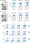
TIM knockdown cells show delayed S phase entry. A and B, left panels, the protein levels of TIM, TIPIN, p21, and p27 were analyzed by SDS-PAGE and Western blotting of whole cell extracts (WCE). HEK293 (A) and U2OS (B) cells were treated with either control or TIM siRNAs. Actin was used as the loading control. Right panels, flow cytometry to determine cell cycle distribution of asynchronized HEK293 (A) and U2OS (B) cells treated with control or TIM siRNAs. The cells were labeled with BrdU and co-stained for BrdU incorporation (y axis) and DNA content (propidium iodide, x axis). Percentages of cells in S phase are shown in black, and the differences in the percentages between control and TIM knockdown cells are shown in red. C, flow cytometry analysis of HEK293 cells treated with control or TIM siRNAs after release from nocodazole blocking at the indicated time points. 2C and 4C represent cells containing one or two copies of each chromosome, respectively. All data are representative of a minimum of two independent experiments. All Western blots in each subfigure were from the same lysate or experiment.
FIGURE 2.
FIGURE 2.
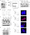
TIM-deficient cells accumulate high levels of replication inhibitors without accumulating DNA damage. A, the protein levels of TIM, TIPIN, p21, and p27 were analyzed by SDS-PAGE and Western blotting of WCE from HEK293T cells, which contain the SV40 large T antigen, treated with either control or TIM siRNAs. Actin was used as the loading control. B, flow cytometry of HEK293T control (left panel) and TIM knockdown (right panel) cells after release from nocodazole block at the indicated time points. C, the protein levels of TIM, p21, p27, SKP2, and cyclin A were analyzed on Western blots of WCEs that were prepared from the synchronized control and TIM knockdown HEK293T cells shown in B. Actin was used as the loading control for SDS-PAGE. noc, nocodazole. D, the protein levels of TIM, CHK1, and p-CHK1 were analyzed by Western blotting using WCE prepared from control and TIM knockdown HEK293T cells with or without UV irradiation. E, quantification of the percentage of HEK293T cells (>200 cells/sample) containing ssDNA, based on positive staining of pulse-labeled BrdU under non-denaturing conditions 2, 6, and 13 h after release from nocodazole treatment. Error bars represent S.D. F—I, flow cytometry (left panels) and representative immunofluorescent staining of the pulse-labeled BrdU (right panels) in control siRNA-treated HEK293T cells (F and G) and TIM siRNA-treated cells (H and I) shown in E. J, the protein levels of TIM, SKP2, and p27 were analyzed by SDS-PAGE and Western blotting of WCE from HEK293T, U2OS, or HT0180 cells treated with either control or TIM siRNAs. Actin was used as the loading control. 2C and 4C represents cell containing one or two copies of each chromosome, respectively. All data are representative of a minimum of two independent experiments. All Western blots in each subfigure were from the same lysate or experiment.
FIGURE 3.
FIGURE 3.
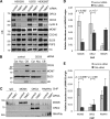
Reduced chromatin recruitment of CDC6 and cyclin E in TIM knockdown cells. A, Western blotting analyses of CB ORC2, CDC6, cyclin E, MCM2-7 (represented by MCM7), CDC45, and GINS (represented by SLD5 and PSF2) in the soluble chromatin fractions isolated from HEK293, HEK293T, and U2OS cells treated either with control or TIM siRNAs. Histone H3 was used as the loading control. B, Western blotting analyses of CDC6, MCM2-7 (represented by MCM7) and GINS (represented by SLD5) in the cytoplasmic (Cyt), nucleoplasmic (Nuc), and CB fractions prepared from HEK293T cells treated with control or CDC6 siRNA. C, Western blotting analysis of MCM5, ORC2, and active RNAPII isolated from ChIP in formaldehyde-cross-linked control and TIM knockdown cells. D and E, real-time PCR analysis of the relative abundance of MCM5, ORC2, and RNAPII bound to the lamin B2 origin in HEK293 cells (D) and HEK293T cells (E) 6 h after nocodazole release, when cells were predominantly in G1 phase. ChIP using IgG alone was used as the background level, and the relative abundance of MCM5, ORC2, and RNAPII shown was after subtracting the background signal from IgG ChIP. Each value represents the mean ± S.D. calculated from triplicate PCRs per one representative experiment. All data are representative of a minimum of two independent experiments. All Western blots in each subfigure were from the same lysate or experiment.
FIGURE 4.
FIGURE 4.
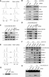
TIM knockdown cells accumulate aberrant CMG helicase complexes. A, top panels, protein levels of TIM, CDC6, and MCM7 were analyzed on Western blots of WCE prepared from U2OS cells stably expressing FLAG-CDC45 and treated with control, TIM, CDC6, or TIM-CDC6 (double knockdown) siRNAs. Bottom panels, Western blotting analyses of FLAG-CDC45 complexes immunopurified (IP) from soluble chromatin fractions isolated from the corresponding knockdown cells to detect the presence of CDC45 (FLAG), MCM2-7 (MCM2 and MCM7), and GINS (SLD5 and PSF2). B, Western blotting analyses of the protein levels of TIM, TIPIN, and CDC6 in WCE (top panels), and the presence of MCM7, SLD5, and MCM10 in the FLAG-CDC45 prepared from HEK293 cells stably expressing FLAG-CDC45 (bottom panels), as performed in (A), with the exception that a lane containing WCE and FLAG-CDC45 IP prepared from TIM-CDC6 double knockdown cells complemented with a siRNA-resistant TIM expression construct was also included. C, flow cytometry analysis of HEK293 cells treated with control, TIM, CDC6, or CDC6-TIM double siRNAs. Percentages of cells in S phase are shown in black, and the differences in the percentages between control and knockdown cells are shown in red. All data are representative of a minimum of two independent experiments. All Western blots in each subfigure were from the same lysate or experiment.
FIGURE 5.
FIGURE 5.
Aberrant CMG helicase complexes accumulate on the chromatin of non-S phase cells depleted with TIM. A, flow cytometry of control cells (left panel) and TIM knockdown HEK293T cells (right panel) stably expressing FLAG-CDC45 at different time points after release from nocodazole block. B, Western blotting analysis of FLAG-CDC45 complexes immunopurified from the soluble chromatin fractions of control and TIM knockdown cells synchronized at different cell cycle stages, as described in A, to detect the presence of CDC45 (FLAG), TIM, MCM2-7 (MCM5 and MCM7), and GINS (SLD5 and PSF2). Noc, nocodazole. C, the protein levels of TIM, CDC6, and SLD5 were analyzed by Western blotting using WCE prepared from control, TIM knockdown, CDC6 knockdown, and TIM-CDC6 double knockdown HEK293T cells expressing FLAG-RECQ4. D, Western blotting analysis of FLAG-RECQ4 complexes immunopurified from the soluble chromatin fractions of the corresponding knockdown cells described in C using antibodies against FLAG, MCM10, MCM7, CDC45, and SLD5. E, flow cytometry of control cells (left panel) and TIM knockdown cells (right panel) stably expressing FLAG-RECQ4 at multiple time points after release from nocodazole block. F, Western blotting analysis of FLAG-RECQ4 complexes immunopurified from the soluble chromatin fractions of control and TIM knockdown cells synchronized at various cell cycle stages, as described in E, to detect the presence of RECQ4 (FLAG), TIPIN, MCM10, MCM2-7 (MCM7), CDC45 and GINS (SLD5 and PSF2). G, Western blotting analysis of FLAG-RECQ4 complexes immunopurified from the chromatin fractions prepared from control cells and TIM siRNA knockdown cells using anti-FLAG antibody. H, helicase activity of equal amounts of FLAG-RECQ4 complex purified from control cells or TIM knockdown HEK293T cells assayed using 32P-labeled splayed-arm substrates. The 32P-labeled ssDNA products were visualized by autoradiography following neutral PAGE. All data are representative of a minimum of three independent experiments. All Western blots in each subfigure were from the same lysate or experiment. 2C and 4C represents cell containing one or two copies of each chromosome, respectively.
FIGURE 6.
FIGURE 6.
The aberrant CMG helicase complexes in TIM knockdown cells exhibit altered posttranslational modifications. A, Western blotting analysis of chromatin-bound FLAG-MCM4 purified from HEK293T cells treated with DMSO, CDC7 inhibitor (PHA 767491), or CDK inhibitor (roscovitine). B, Western blotting analysis of TIM, MCM2, MCM4, and phosphorylated polypeptides in chromatin-bound FLAG-MCM7 complexes purified from control and TIM knockdown cells. C, Western blotting analysis of MCM4, MCM7, and MCM2 of the chromatin-bound FLAG-MCM7 complex purified from control or TIM knockdown HEK293T cells treated with DMSO or APH. D, amino acid sequence of the first 150 residues of human MCM4. The three intrinsic DDK target sites (S/T-D/E) are underlined, and the six PG sites (S/T-S/T-P) are shown in bold. E, Western blotting analysis of chromatin-bound FLAG-MCM4 WT, the phosphomimetic DDK mutant (3E-D/E), and the phosphomimetic PG mutant (6E-EP) purified from control and TIM knockdown cells. F, Western blotting analysis of chromatin-bound FLAG-MCM4 WT and phosphodefective PG mutant (6A-AP) purified from control and TIM knockdown cells. G, Western blotting analysis of MCM4 in purified FLAG-CDC45 or FLAG-MCM7 complexes treated with or without λ phosphatase (λ PPase). FLAG-CDC45 and FLAG-MCM7 were detected using an anti-FLAG antibody. H, Western blotting analysis of chromatin-bound FLAG-CDC45 complexes purified from HEK293 cells (left two lanes) or U2OS cells (right two lanes) treated with control or TIM siRNA. I, Western blotting analysis of chromatin-bound FLAG-CDC45 complexes purified from control or TIM siRNA-treated HEK293T cells. The purified complexes were analyzed using antibodies to detect MCM2-7 (MCM4, MCM2), GINS (SLD5), CDC45 (FLAG), and DNA polymerase δ (Pol δ). All data are representative of a minimum of three independent experiments. All Western blots in each subfigure were from the same lysate or experiment.
FIGURE 7.
FIGURE 7.
TIM-TIPIN associates with MCM2-7 prior to DNA replication initiation. A, Western blotting analysis for the presence of TIM, TIPIN, MCM2-7 (MCM7), and CDC6 in FLAG complexes purified from the cytoplasmic (cyt), nucleoplasmic (nuc), and CB fractions prepared from HEK293T cells transfected with a control vector or a vector expressing FLAG-TIM. B, flow cytometry of HEK293T cells stably expressing FLAG-MCM7 at the indicated time points after release from nocodazole block. C, Western blotting analysis of FLAG-MCM7 complexes immunopurified with anti-FLAG M2-agarose from soluble chromatin-bound fractions of HEK293T cells synchronized at different time points after nocodazole (Noc) release as described in B. The blots were probed with the indicated antibodies to detect the presence of MCM2-7 (MCM4 and FLAG), GINS (PSF2 and SLD5), CDC45, TIM, and TIPIN. D, flow cytometry of HEK293T cells stably expressing FLAG-CDC45 at the indicated time points after release from nocodazole block. E, Western blotting analysis of the FLAG-CDC45 complex immunopurified with anti-FLAG M2-agarose from soluble chromatin-bound fractions of HEK293T cells synchronized at different time points after nocodazole release as described in D. The purified complexes were analyzed with the indicated antibodies to detect the presence of MCM2-7 (MCM4 and MCM7), GINS (PSF2 and SLD5), and TIM. F, the protein levels of RECQ4, MCM10, and CDC6 were analyzed by Western blotting using WCE prepared from control, RECQ4, MCM10, and CDC6 knockdown cells stably expressing FLAG-TIM. G, Western blotting analysis of FLAG-TIM complexes immunopurified from the soluble chromatin fractions of the corresponding knockdown cells described in A to detect the presence of TIM (FLAG), TIPIN, MCM2-7 (MCM7), MCM10, and RECQ4. All data are representatives of a minimum of two independent experiments. All Western blots in each subfigure were from the same lysate or experiment. 2C and 4C represents cell containing one or two copies of each chromosome, respectively.
FIGURE 8.
FIGURE 8.
Model for TIM-TIPIN function in facilitating efficient DNA replication initiation by suppressing chromatin accumulation of aberrant CMG complexes. In normal cells, TIM-TIPIN is known to travel with the CMG complex to maintain proper replication fork progression (left). However, TIM-TIPIN also interacts with MCM2-7 outside of S phase. We suggest that, after completion of DNA synthesis, the interaction of TIM-TIPIN with MCM2-7 may be responsible for the disassembly and dissociation of the CMG from the chromatin prior to mitosis or entry to the next round of the cell cycle. In the absence of TIM, aberrant CMG complexes are present on the chromatin during G1 phase (right). The presence of these abnormal CMG complexes may provide false signals to prevent CDC6-dependent CMG assembly at active origins and initiate DNA replication.

Similar articles

Cited by

References

    1. Dimitrova D. S., Todorov I. T., Melendy T., and Gilbert D. M. (1999) Mcm2, but not RPA, is a component of the mammalian early G1-phase prereplication complex. J. Cell Biol. 146, 709–722 - PMC - PubMed
    1. Edwards M. C., Tutter A. V., Cvetic C., Gilbert C. H., Prokhorova T. A., and Walter J. C. (2002) MCM2-7 complexes bind chromatin in a distributed pattern surrounding the origin recognition complex in Xenopus egg extracts. J. Biol. Chem. 277, 33049–33057 - PubMed
    1. Woodward A. M., Göhler T., Luciani M. G., Oehlmann M., Ge X., Gartner A., Jackson D. A., and Blow J. J. (2006) Excess Mcm2-7 license dormant origins of replication that can be used under conditions of replicative stress. J. Cell Biol. 173, 673–683 - PMC - PubMed
    1. Kim J., and Kipreos E. T. (2008) Control of the Cdc6 replication licensing factor in metazoa: the role of nuclear export and the CUL4 ubiquitin ligase. Cell Cycle 7, 146–150 - PubMed
    1. Labib K. (2010) How do Cdc7 and cyclin-dependent kinases trigger the initiation of chromosome replication in eukaryotic cells? Genes Dev. 24, 1208–1219 - PMC - PubMed

MeSH terms

LinkOut - more resources