Comparative Study
. 2016 Sep 27;134(13):934-44.
doi: 10.1161/CIRCULATIONAHA.116.022684. Epub 2016 Sep 1.

Percutaneous Device Closure of Paravalvular Leak: Combined Experience From the United Kingdom and Ireland

Affiliations
Comparative Study

Percutaneous Device Closure of Paravalvular Leak: Combined Experience From the United Kingdom and Ireland

Patrick A Calvert et al. Circulation. .

Erratum in

Abstract

Background: Paravalvular leak (PVL) occurs in 5% to 17% of patients following surgical valve replacement. Percutaneous device closure represents an alternative to repeat surgery.

Methods: All UK and Ireland centers undertaking percutaneous PVL closure submitted data to the UK PVL Registry. Data were analyzed for association with death and major adverse cardiovascular events (MACE) at follow-up.

Results: Three hundred eight PVL closure procedures were attempted in 259 patients in 20 centers (2004-2015). Patient age was 67±13 years; 28% were female. The main indications for closure were heart failure (80%) and hemolysis (16%). Devices were successfully implanted in 91% of patients, via radial (7%), femoral arterial (52%), femoral venous (33%), and apical (7%) approaches. Nineteen percent of patients required repeat procedures. The target valve was mitral (44%), aortic (48%), both (2%), pulmonic (0.4%), or transcatheter aortic valve replacement (5%). Preprocedural leak was severe (61%), moderate (34%), or mild (5.7%) and was multiple in 37%. PVL improved postprocedure (P<0.001) and was none (33.3%), mild (41.4%), moderate (18.6%), or severe (6.7%) at last follow-up. Mean New York Heart Association class improved from 2.7±0.8 preprocedure to 1.6±0.8 (P<0.001) after a median follow-up of 110 (7-452) days. Hospital mortality was 2.9% (elective), 6.8% (in-hospital urgent), and 50% (emergency) (P<0.001). MACE during follow-up included death (16%), valve surgery (6%), late device embolization (0.4%), and new hemolysis requiring transfusion (1.6%). Mitral PVL was associated with higher MACE (hazard ratio [HR], 1.83; P=0.011). Factors independently associated with death were the degree of persisting leak (HR, 2.87; P=0.037), New York Heart Association class (HR, 2.00; P=0.015) at follow-up and baseline creatinine (HR, 8.19; P=0.001). The only factor independently associated with MACE was the degree of persisting leak at follow-up (HR, 3.01; P=0.002).

Conclusion: Percutaneous closure of PVL is an effective procedure that improves PVL severity and symptoms. Severity of persisting leak at follow-up is independently associated with both MACE and death. Percutaneous closure should be considered as an alternative to repeat surgery.

Keywords: catheters; heart failure; paravalvular regurgitation; survival.


Figures

Figure 1
Figure 1. 3D-TEE images of bileaflet mechanical mitral valve replacement (MVR).
(A) 3D-TEE ”surgical view” of the MVR viewed from the left atrial side. All the other panels are aligned in the same orientation. This view is used to guide crossing the mitral PVL with guidewire and delivery sheath from a transseptal puncture. The MVR sewing ring is indicated by the large arrow head in all panels. The MVR is seen with the leaflets in the open position (small arrows). Ao (aortic rim), LAA (left atrial appendage rim), IAS (inter-atrial septal rim), post (posterior rim), lat (lateral rim). (B) Agilis steerable sheath (SJM, Plymouth, MN) (thin arrow) with deflectable tip with acute angulation in order to cross a PVL in the IAS rim. (C) PVL in the LAA rim with a guidewire across it (thin arrow). (D) PVL in the postero-septal rim with a device deployed in it (thin arrow).
Figure 2
Figure 2. TEE and fluoroscopic images of PVLs of aortic and mitral valve replacements during device closure
(A) TEE image of a PVL of an aortic valve replacement (AVR) with the perimeter traced with the dotted line. Note the crescentic shape. The image cross-section is just out of plane of the surgical valve leaflets. (B) TEE image of a guidewire (white arrow) crossing a PVL of an AVR (red arrowhead). (C) Fluoroscopic image of an AVP3 device (white arrow) deployed across an AVR (red arrowhead) but still on the delivery cable. (D) Same device as panel C with the device now released. Each disc is highlighted with a white arrow. Note the device conforms to the shape of the PVL. (E and F) Fluoroscopic image of an AVP3 device deployed across a PVL of an MVR (white arrowhead). The device is still on the delivery cable. Note the patient also has an AVR (red arrowhead). Each of the discs of the device is highlighted with a white arrow. Note that the device has shaped up well suggesting the device is not oversized. In Panel E the orientation of the image is cross-sectional to the MVR which is good for deploying the device’s discs. In Panel F the orientation of the image is ’en face’ to the MVR which is good for ensuring that the device’s discs do not interfer with the movement of the MVR leaflets.
Figure 3
Figure 3
The six most frequent devices used for paravalvular leak closure with frequencies. AVP (Amplatzer™ Vascular Plug, St Jude Medical [SJM], Plymouth, MN), ADO (Amplatzer™ Duct Occluder, SJM, Plymouth, MN), mVSD (muscular Ventricular Septal Defect Occluder, SJM, Plymouth, MN), PLD (Paravalvular Leak Device, Occlutech®, Jena, Germany). AVP3 and PLD were not available for the entire duration of the study and are not currently FDA approved. Images reproduced with permission of SJM and Occlutech®
Figure 4
Figure 4. Univariable analysis for variables associated with death on follow up.
Data plotted are unadjusted hazard ratio (HR) (box) and 95% confidence interval (stems). y (years), LV (left ventricle), NYHA (New York Heart Association), MVR (mitral valve replacement), std dev (standard deviation). For definitions of HR comparisons please see statistical methodology. *Factors entered into multivariable analysis.
Figure 5
Figure 5. Univariable analysis for variables associated with MACE on follow up.
Data plotted are unadjusted hazard ratio (HR) (box) and 95% confidence interval (stems). MACE (major adverse cardiovascular event, y (years), LV (left ventricle), NYHA (New York Heart Association) , MVR (mitral valve replacement), std dev (standard deviation). For definitions of HR comparisons please see statistical methodology. *Factors entered into multivariable analysis.
Figure 6
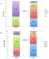
Figure 6. (A) Severity of Paravalvular Leak and (B) New York Heart Association Functional Classification before and after percutaneous closure attempt
Figure 7
Figure 7. Kaplan-Meier cumulative survival plots according to the severity of the residual PVL leak at follow up
Figure 8
Figure 8. Kaplan-Meier cumulative “survival free from MACE” plots according to the severity of the residual PVL leak at follow up
MACE (major adverse cardiovascular event)

References

    1. Herr R, Starr A, Mccord CW, Wood JA. SPECIAL PROBLEMS FOLLOWING VALVE REPLACEMENT: EMBOLUS, LEAK, INFECTION, RED CELL DAMAGE. Ann Thorac Surg. 1965;1:403–415. - PubMed
    1. Hammermeister K, Sethi GK, Henderson WG, Grover FL, Oprian C, Rahimtoola SH. Outcomes 15 years after valve replacement with a mechanical versus a bioprosthetic valve: final report of the Veterans Affairs randomized trial. J Am Coll Cardiol. 2000;36:1152–1158. - PubMed
    1. Ionescu A, Fraser AG, Butchart EG. Prevalence and clinical significance of incidental paraprosthetic valvar regurgitation: a prospective study using transoesophageal echocardiography. Heart Br Card Soc. 2003;89:1316–1321. - PMC - PubMed
    1. Gafoor S, Steinberg DH, Franke J, Bertog S, Vaskelyte L, Hofmann I, Sievert H. Tools and Techniques - Clinical: Paravalvular leak closure. EuroIntervention. 2014;9:1359–1363. - PubMed
    1. Rihal CS, Sorajja P, Booker JD, Hagler DJ, Cabalka AK. Principles of percutaneous paravalvular leak closure. JACC Cardiovasc Interv. 2012;5:121–130. - PubMed

Publication types