Review
. 2016 Sep;7(5):561-77.
doi: 10.1177/2150135116651114.

Development and Morphology of the Ventricular Outflow Tracts

Affiliations
Review

Development and Morphology of the Ventricular Outflow Tracts

Robert H Anderson et al. World J Pediatr Congenit Heart Surg. 2016 Sep.

Abstract

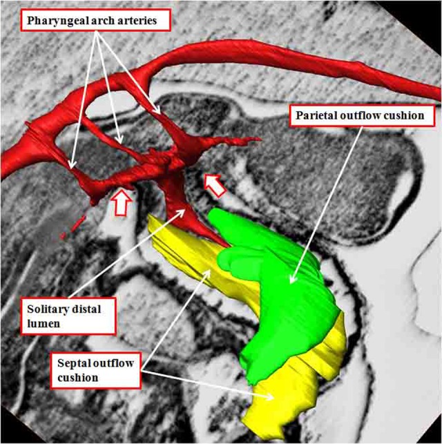
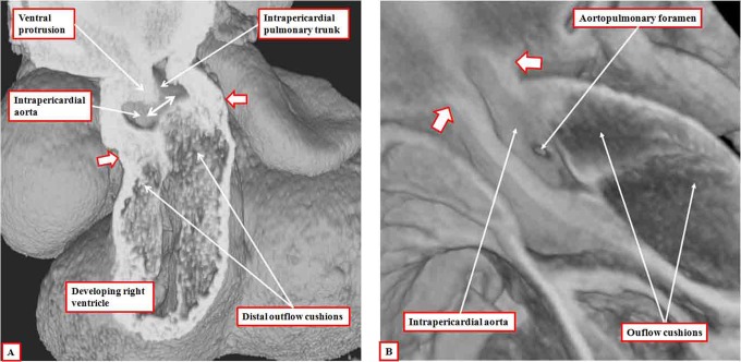
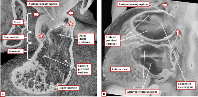
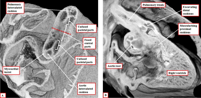
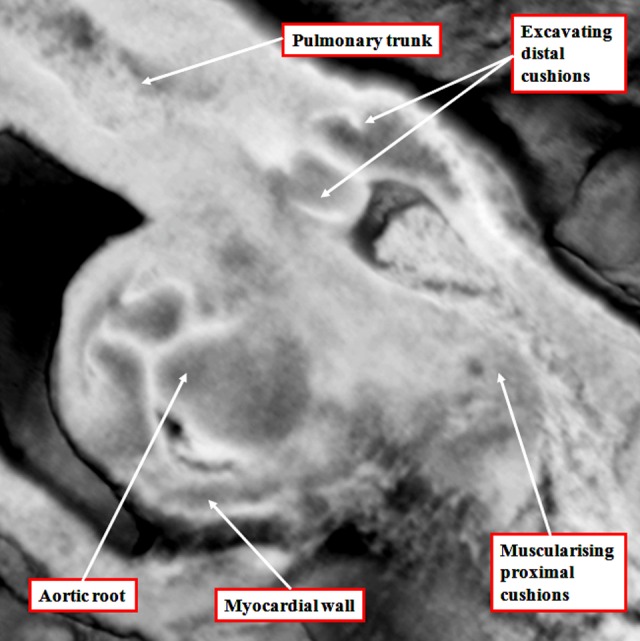
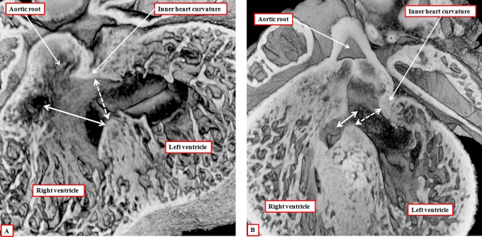
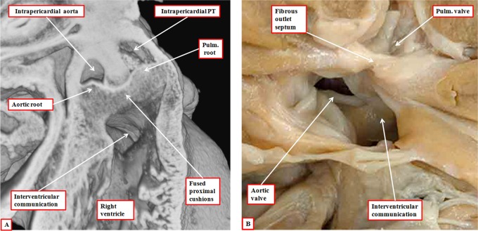
It is customary, at the current time, to consider many, if not most, of the lesions involving the ventricular outflow tract in terms of conotruncal malformations. This reflects the introduction, in the early 1940s, of the terms conus and truncus to describe the components of the developing outflow tract. The definitive outflow tracts in the postnatal heart, however, possess three, rather than two, components. These are the intrapericardial arterial trunks, the arterial roots, and the subvalvar ventricular outflow tracts. Congenital lesions afflicting the arterial roots, however, are not currently considered to be conotruncal malformations. This suggests a lack of logic in the description of cardiac development and its use as a means of categorizing congenital malformations. It is our belief that the developing outflow tract, like the postnatal outflow tracts, can readily be described in tripartite fashion, with its distal, intermediate, and proximal components forming the primordiums of the postnatal parts. In this review, we present evidence obtained from developing mice and human hearts to substantiate this notion. We show that the outflow tract, initially with a common lumen, is divided into its aortic and pulmonary components by a combination of an aortopulmonary septum derived from the dorsal wall of the aortic sac and outflow tract cushions that spiral through its intermediate and proximal components. These embryonic septal structures, however, subsequently lose their septal functions as the outflow tracts develop their own discrete walls. We then compare the developmental findings with the anatomic arrangements seen postnatally in the normal human heart. We show how correlations with the embryologic findings permit logical analysis of the congenital lesions involving the outflow tracts.

Keywords: conotruncal anomalies; conus; episcopic microscopy; multidetector computer tomography; normal anatomy; truncus.


Conflict of interest statement

The author(s) declared no potential conflicts of interest with respect to the research, authorship, and/or publication of this article.

Figures

Figure 1.
Figure 1.
The images, prepared using dissection and scanning electron microscopy, show the developing heart tube as seen in the mouse early (panel A) and late (panel B) during the ninth day of embryonic development. This is equivalent of around five weeks of development in man.
Figure 2.
Figure 2.
The images are from an episcopic data set prepared from a human embryo at Carnegie stage 14, representing the end of the fifth week of development. The left panel (A) shows the dog-leg bend in the outflow tract, which at this early stage is supported exclusively from the developing right ventricle. The right panel (B), cut in frontal fashion at the junction of the outflow tract with the pharyngeal mesenchyme, shows the origins of the pharyngeal arch arteries from the aortic sac. The back wall of the sac, shown by the star, is the putative aortopulmonary septum. The two single-headed arrows show the extent of the pericardial cavity.
Figure 3.
Figure 3.
The image shows the outflow cushions reconstructed from an episcopic data set prepared from a mouse embryo killed early during the 12th day of development. The outflow cushions, shown in green and yellow, no longer reach the margins of the pericardial cavity (white arrows with red borders). The solitary lumen of the distal outflow tract becomes continuous dorsally with the cavity of the aortic sac, from which arise the pharyngeal arch arteries, at this stage bilaterally symmetrical.
Figure 4.
Figure 4.
The images are both made using episcopic data sets from mice embryos killed toward the end of the 12th day of development. The left panel (A) shows a frontal section revealing the oblique nature of the ventral protrusion, which is the aortopulmonary septum. At this stage, there is an aortopulmonary foramen (double-headed white arrow) between the proximal end of the protrusion and the fused distal end of the outflow cushions. The white arrows with dark borders show the distal margin of the myocardium, which now encloses only the intermediate and proximal parts of the outflow tract. Panel B, from a different data set, shows the aortopulmonary foramen as seen from the cavity of the aorta, having cut away the parietal aortic wall. The white arrows with dark borders in this panel show the distal extent of the pericardial cavity.
Figure 5.
Figure 5.
Panel A is prepared using an episcopic data set from a mouse embryo killed at the end of the 12th day of development. It shows how the aortopulmonary septum, formed by the protrusion from the dorsal wall of the aortic sac, has fused with the distal ends of the outflow cushions (dashed white line). The white arrows with dark borders show the extent of the pericardial cavity. The distal outflow tract is fully divided at this stage. The fused distal cushions have also divided the intermediate part of the outflow tract. Note the appearance of the intercalated cushions (white stars with dark borders) in this middle component. The outflow cushions, however, remain unfused in the proximal part of the outflow tract. Panel B is from an episcopic data set prepared from a human embryo at Carnegie stage 16. It shows how columns of condensed mesenchyme, derived from the cells migrating from the neural crest, occupy the fused distal and the unfused proximal parts of the outflow cushions. They are not involved with separating the intrapericardial arterial trunks, which have already been separated by the aortopulmonary septum, formed from the protrusion of the dorsal wall of the aortic sac.
Figure 6.
Figure 6.
The left panel (A) is prepared using an episcopic data set from a mouse embryo killed during the 13th day of development. The short axis of the intermediate part of the outflow tract is viewed from above, showing the developing primordiums of the aortic root. Note that, at this stage, the roots remained encased in a turret of outflow tract myocardium. The right panel (B) is from an episcopic data set prepared using a human embryo at Carnegie stage 20. The three parts of the outflow tract are shown, with the distal cushions excavating to form the leaflets of the pulmonary valve, and the surface of the fused proximal cushions muscularizing to form the subpulmonary infundibulum. At this stage, the intermediate and proximal parts of the outflow tract retain their myocardial walls.
Figure 7.
Figure 7.
The image, a magnification of the section shown as Figure 6B, reveals the start of the excavation of the cushions that will eventually produce the leaflets of the arterial valves. At this stage, the intermediate component of the outflow tract remains encased within a myocardial turret.
Figure 8.
Figure 8.
The images are from episcopic data sets prepared from developing mice killed during the 13th embryonic day (panel A) and at the beginning of the 14th day of development (panel B). They show that, as the aortic root is transferred so as to take origin from the left as opposed to the right ventricle, the plane of the initial interventricular communication (dashed double-headed white arrow) becomes the left ventricular outflow tract. This plane is roofed by the inner heart curvature. The plane from the ventricular septal crest to the fused proximal outflow cushions (double-headed solid white arrow) then becomes the interventricular communication. Note that, at this stage, the distal myocardial boundary remains confluent with the edges of the excavating distal cushions.
Figure 9.
Figure 9.
The images are from different episcopic data sets prepared from mice killed during the 15th day of embryonic development. They show, in the left panel (A), how the rightward margins of the atrioventricular (AV) cushions close the persisting interventricular communication. The right panel shows how, subsequent to closure of the foramen, the inner heart curvature remains interposed between the developing leaflets of the aortic and mitral valves. Note the developing sinuses of the aortic root and the three components of the left ventricular outflow tract.
Figure 10.
Figure 10.
The image is prepared from a data set obtained using multidetector computed tomography in a patient undergoing investigation for coronary arterial disease. The pericardial reflections divide the ascending aorta into intrapericardial and extrapericardial components. The pulmonary trunk branches at the margins of the pericardial cavity into the right and left pulmonary arteries.
Figure 11.
Figure 11.
The virtual dissections are made from a data set obtained using multidetector computed tomography in a patient undergoing investigation for coronary arterial disease. They show how each outflow tract is formed in a tripartite fashion, with the components represented by the intrapericardial arterial trunk, the arterial root, and the subvalvar outflow tract, respectively. Note that, in the right ventricle (panel A), the subvalvar component is a completely muscular infundibular sleeve, whereas in the left ventricle (panel B), the posterior wall of the subvalvar area is formed by fibrous continuity between the leaflets of the aortic and mitral valves.
Figure 12.
Figure 12.
The reconstruction, seen in short-axis projection from the cardiac apex, shows the spiraling nature of the intrapericardial arterial trunks. The aorta is shown in red, and the pulmonary trunk in blue.
Figure 13.
Figure 13.
The virtual dissections show how the pulmonary (panel A) and aortic (panel B) roots are limited distally by the sinotubular junctions (solid double-headed arrow) but proximally by a virtual plane made by joining together the basal attachments of the semilunar leaflets (double-headed dashed arrow). Note that the virtual basal plane is proximal to the anatomic ventriculoarterial junctions (white arrows with dark borders).
Figure 14.
Figure 14.
The three-dimensional data set made available by means of multidetector computed tomography from a patient undergoing investigation of coronary arterial disease has been segmented and reconstructed to show the hinges of the leaflets of the arterial and atrioventricular valves. The leaflets of the atrioventricular valves are hinged in an annular fashion, whereas the hinges of the arterial valves, when reconstructed, take the form of three-pointed coronets.
Figure 15.
Figure 15.
The outflow tract of the normal heart has been opened through an incision across the left coronary sinus of the aortic root. The membranous septum has been transilluminated from the right side. This shows how the triangular space between the right coronary and the nonadjacent sinuses of the root is filled by a fibrous wall. Similar triangles with fibrous walls are to be found beneath each of the valvar commissures, these being the points at which the leaflets join together at the sinotubular junction (white stars with dark borders). The relationships of these triangles are shown in Figure 16. Note how the virtual basal plane is created by joining together the basal attachments of the valvar leaflets (dotted line).
Figure 16.
Figure 16.
Virtual dissection of the aortic root shows how the tips of each of the interleaflet triangles (white arrows with dark borders) point outside the cardiac cavities. Panel A shows a cut through the triangle between the right and left coronary aortic sinuses and is viewed from behind. Panel B shows a cut across the triangle between the right coronary and the nonadjacent aortic sinuses and is shown from the front. Note that the membranous septum forms the base of this triangle (see also Figure 15). Panel C, showing a cut through the triangle between the left coronary and the nonadjacent aortic sinuses, is viewed from the left side.
Figure 17.
Figure 17.
The images show virtual dissections revealing the structure of the right ventricular infundibulum. The left panel (A) is made by cutting away the parietal wall of the right ventricle, showing how the posterior wall of the infundibulum is formed by the supraventricular crest. The larger part of this wall is no more than the inner heart curvature. The component adjacent to the aortic root, however, is the free-standing infundibular sleeve, the presence of which makes it possible to remove the root for use as an autograft in the Ross procedure. The right panel (B) shows this sleeve, revealing the tissue place that separates it from the aortic root.
Figure 18.
Figure 18.
The images show an aortopulmonary window (panel A), with origin of the right pulmonary artery on the aortic side of the window (panel B). This is well explained by inadequate growth of the aortopulmonary septum from the dorsal wall of the aortic sac (see Figure 4A), leaving a fold between the walls of the aorta and the pulmonary trunk at the margins of the pericardial cavity (star in panel C). Note the separate formation of the aortic and pulmonary roots, implying normal septation and separation of the intermediate and proximal components of the developing outflow tract.
Figure 19.
Figure 19.
The images are from embryonic mice in which the gene for Tbx1 has been knocked out. All these mice develop with common arterial trunks. The images are from embryos killed on the 13th day of development. The left panel (A) shows the common trunk, feeding the systemic and pulmonary arteries, with no formation of the aortopulmonary septum. It also shows failure of fusion of the outflow cushions. The right panel, from a different embryo at the same stage of development, shows a cross section through the intermediate part of the outflow tract. Both the intercalated cushions are seen, together with the distal ends of the unfused major outflow cushions. This template provides the primordiums for the formation of a common truncal valve with four leaflets. Note that the cushions are contained within the turret of myocardium that surrounds the intermediate part of the outflow tract.
Figure 20.
Figure 20.
In panel A, we show the features of the bicuspid aortic valve with the conjoined leaflet derived from the leaflets that usually guard the valvar sinuses giving rise to the coronary arteries. There is formation only of the interleaflet triangles between the nonadjacent leaflet and the conjoined leaflet (white arrows with dark borders), with a raphe formed at the anticipated site of the interleaflet triangle between the two coronary aortic sinuses. Panel B shows reconstruction of the hinges of the valvar leaflets in an adult with a bicuspid aortic valve and conjunction of the leaflets guarding the coronary aortic sinuses. The anticipated point of the coronet at the site of the fused leaflets does not extend to the sinotubular junction (long double-headed arrow), in contrast to the zones of apposition with the other leaflet, which extend to the sinotubular junction (short double-headed arrows).
Figure 21.
Figure 21.
The left panel (A) is taken from an episcopic data set prepared from a developing mouse embryo at the 15th day of development with knockout of the Furin enzyme. The aortopulmonary septum has fused with the distal cushions to produce separate intrapericardial arterial trunks. The outflow cushions themselves are also fused, but with minimal development of the proximal components, which have failed to muscularize. As a result, the arterial trunks remain supported by the right ventricle, the interventricular communication being doubly committed. This arrangement is seen postnatally in the heart with double outlet right ventricle shown in the right panel (B). It has a fibrous rather than a muscular outlet septum. The interventricular communication, opening to the right ventricle between the limbs of the septomarginal trabeculation, is doubly committed.

References

    1. Kramer TC. The partitioning of the truncus and conus and the formation of the membranous portion of the interventricular septum in the human heart. Am J Anat. 1942;71(3):343–370.
    1. Roberts WC. The 2 most common congenital heart diseases. Am J Cardiol. 1984;53(8):1198. - PubMed
    1. Lacour Gayet F, Bove EL, Hraska V, Morell VO, Spray TL. Surgery of Conotruncal Anomalies. Switzerland: Springer International Publishing; 2016.
    1. Sizarov A, Lamers WH, Mohun TJ, Brown NA, Anderson RH, Moorman AF. Three-dimensional and molecular analysis of the arterial pole of the developing human heart. J Anat. 2012;220(4):336–349. - PMC - PubMed
    1. Anderson RH, Chaudhry B, Mohun TJ, et al. Normal and abnormal development of the intrapericardial arterial trunks in humans and mice. Cardiovasc Res. 2012;95(1):108–115. - PMC - PubMed

LinkOut - more resources