Case Reports
. 2016 Sep 1:2016:bcr2016216453.
doi: 10.1136/bcr-2016-216453.

OCT angiography in the management of choroidal neovascular membrane secondary to Sorsby fundus dystrophy

Affiliations
Case Reports

OCT angiography in the management of choroidal neovascular membrane secondary to Sorsby fundus dystrophy

Aditi Mohla et al. BMJ Case Rep. .

Abstract

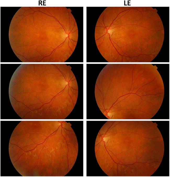
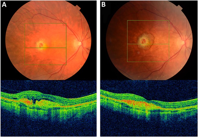
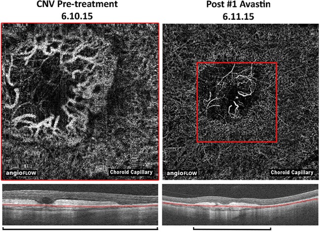
We describe the management of a woman aged 52 years with molecularly confirmed Sorsby fundus dystrophy, who presented with acute visual deterioration in her right eye. Fundus examination identified a right macular lesion suggestive of a choroidal neovascular membrane (CNVM). Optical coherence tomography angiography (OCTA) confirmed the presence of a CNVM. She was treated with 2 monthly intravitreal injections of bevacizumab, associated with OCTA evidence of regression of the CNVM and improvement in her visual acuity. OCTA is a novel, non-invasive method of imaging the retinal vasculature. Images are acquired rapidly, with no associated side effects, offering advantages over the current gold standard technique-fundus fluorescein angiography.


Figures

Figure 1
Figure 1
Fundus photos of Sorsby fundus dystrophy (prechoroidal neovascular membrane).
Figure 2
Figure 2
(A) Fundus photo and corresponding optical coherence tomography scan of right eye choroidal neovascular membrane secondary to Sorsby fundus dystrophy (Pre-treatment). (B) Corresponding images post Intravitreal Bevacizumab #1.
Figure 3
Figure 3
Optical coherence tomography (OCT) angiography images of the choroidal neovascular membrane pretreatment and postbevacizumab treatment, with comparison on OCT (automated segmentation, note the magnification bars).

References

    1. Dansingani KK, Naysan J, Freund KB. En face OCT angiography demonstrates flow in early type 3 neovascularization (retinal angiomatous proliferation). Eye (Lond) 2015;29:703–6. - PMC - PubMed
    1. De Carlo TE, Bonini Filho MA, Chin AT et al. . Spectral-domain optical coherence tomography angiography of choroidal neovascularization. Ophthalmology 2015;122:1228–38. 10.1016/j.ophtha.2015.01.029 - DOI - PubMed
    1. Ishibazawa A, Nagaoka T, Takahashi A et al. . Optical coherence tomography angiography in diabetic retinopathy: a prospective pilot study. Am J Ophthalmol 2015;160:35–44.e1. 10.1016/j.ajo.2015.04.021 - DOI - PubMed
    1. Jia Y, Bailey ST, Hwang TS et al. . Quantitative optical coherence tomography angiography of vascular abnormalities in the living human eye. Proc Natl Acad Sci USA 2015;112:E2395–402. - PMC - PubMed
    1. Spaide RF. Optical coherence tomography angiography signs of vascular abnormalization with antiangiogenic therapy for choroidal neovascularization. Am J Ophthalmol 2015;160:6–16. 10.1016/j.ajo.2015.04.012 - DOI - PubMed

Publication types

MeSH terms

Supplementary concepts