

Diagnosing Lung Nodules on Oncologic MR/PET Imaging: Comparison of Fast T1-Weighted Sequences and Influence of Image Acquisition in Inspiration and Expiration Breath-Hold
- PMID: 27587957
- PMCID: PMC5007395
- DOI: 10.3348/kjr.2016.17.5.684
Diagnosing Lung Nodules on Oncologic MR/PET Imaging: Comparison of Fast T1-Weighted Sequences and Influence of Image Acquisition in Inspiration and Expiration Breath-Hold
Abstract
Objective: First, to investigate the diagnostic performance of fast T1-weighted sequences for lung nodule evaluation in oncologic magnetic resonance (MR)/positron emission tomography (PET). Second, to evaluate the influence of image acquisition in inspiration and expiration breath-hold on diagnostic performance.
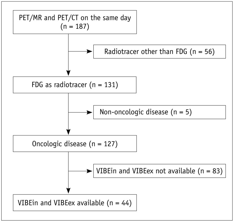
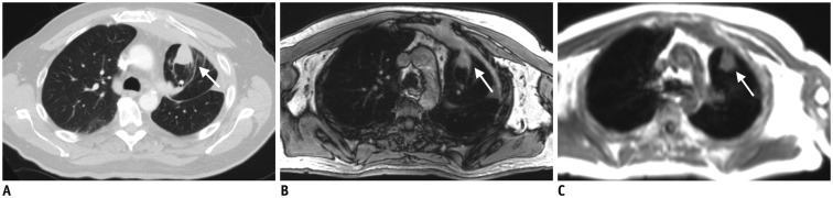
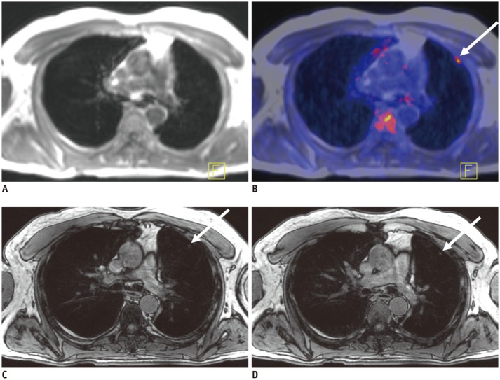
Materials and methods: The study was approved by the local Institutional Review Board. PET/CT and MR/PET of 44 cancer patients were evaluated by 2 readers. PET/CT included lung computed tomography (CT) scans in inspiration and expiration (CTin, CTex). MR/PET included Dixon sequence for attenuation correction and fast T1-weighted volumetric interpolated breath-hold examination (VIBE) sequences (volume interpolated breath-hold examination acquired in inspiration [VIBEin], volume interpolated breath-hold examination acquired in expiration [VIBEex]). Diagnostic performance was analyzed for lesion-, lobe-, and size-dependence. Diagnostic confidence was evaluated (4-point Likert-scale; 1 = high). Jackknife alternative free-response receiver-operating characteristic (JAFROC) analysis was performed.
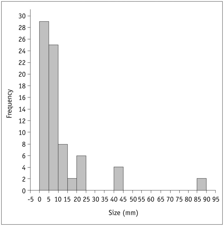
Results: Seventy-six pulmonary lesions were evaluated. Lesion-based detection rates were: CTex, 77.6%; VIBEin, 53.3%; VIBEex, 51.3%; and Dixon, 22.4%. Lobe-based detection rates were: CTex, 89.6%; VIBEin, 58.3%; VIBEex, 60.4%; and Dixon, 31.3%. In contrast to CT, inspiration versus expiration did not alter diagnostic performance in VIBE sequences. Diagnostic confidence was best for VIBEin and CTex and decreased in VIBEex and Dixon (1.2 ± 0.6; 1.2 ± 0.7; 1.5 ± 0.9; 1.7 ± 1.1, respectively). The JAFROC figure-of-merit of Dixon was significantly lower. All patients with malignant lesions were identified by CTex, VIBEin, and VIBEex, while 3 patients were false-negative in Dixon.
Conclusion: Fast T1-weighted VIBE sequences allow for identification of patients with malignant pulmonary lesions. The Dixon sequence is not recommended for lung nodule evaluation in oncologic MR/PET patients. In contrast to CT, inspiration versus expiratory breath-hold in VIBE sequences was less crucial for lung nodule evaluation but was important for diagnostic confidence.
Keywords: Expiration; Inspiration; Lung; MR/PET; PET/MR; PET/MRI; Pulmonary nodule.
Figures
Similar articles
-
PET/MR imaging in the detection and characterization of pulmonary lesions: technical and diagnostic evaluation in comparison to PET/CT.J Nucl Med. 2014 May;55(5):724-9. doi: 10.2967/jnumed.113.129247. Epub 2014 Mar 20. J Nucl Med. 2014. PMID: 24652827
-
Performance of whole-body integrated 18F-FDG PET/MR in comparison to PET/CT for evaluation of malignant bone lesions.J Nucl Med. 2014 Feb;55(2):191-7. doi: 10.2967/jnumed.113.123646. Epub 2013 Dec 5. J Nucl Med. 2014. PMID: 24309383
-
Pulmonary nodule detection in oncological patients - Value of respiratory-triggered, periodically rotated overlapping parallel T2-weighted imaging evaluated with PET/CT-MR.Eur J Radiol. 2018 Jan;98:165-170. doi: 10.1016/j.ejrad.2017.11.010. Epub 2017 Nov 16. Eur J Radiol. 2018. PMID: 29279157
-
Improved Detection of Small Pulmonary Nodules Through Simultaneous MR/PET Imaging.PET Clin. 2018 Jan;13(1):89-95. doi: 10.1016/j.cpet.2017.09.001. PET Clin. 2018. PMID: 29157389 Review.
-
Imaging of Osteoarthritis by Conventional Radiography, MR Imaging, PET-Computed Tomography, and PET-MR Imaging.PET Clin. 2019 Jan;14(1):17-29. doi: 10.1016/j.cpet.2018.08.004. Epub 2018 Oct 24. PET Clin. 2019. PMID: 30420217 Review.
Cited by
-
Current Applications for Nuclear Medicine Imaging in Pulmonary Disease.Curr Pulmonol Rep. 2020;9(3):82-95. doi: 10.1007/s13665-020-00251-1. Epub 2020 Jul 22. Curr Pulmonol Rep. 2020. PMID: 32837866 Free PMC article. Review.
-
Evaluation of 68Ga-DOTATOC PET/MRI for whole-body staging of neuroendocrine tumours in comparison with 68Ga-DOTATOC PET/CT.Eur Radiol. 2017 Oct;27(10):4091-4099. doi: 10.1007/s00330-017-4803-2. Epub 2017 Apr 24. Eur Radiol. 2017. PMID: 28439648
-
Use of "Diagnostic Yield" in Imaging Research Reports: Results from Articles Published in Two General Radiology Journals.Korean J Radiol. 2022 Dec;23(12):1290-1300. doi: 10.3348/kjr.2022.0741. Korean J Radiol. 2022. PMID: 36447417 Free PMC article.
-
Detection of lung lesions in breath-hold VIBE and free-breathing Spiral VIBE MRI compared to CT.Insights Imaging. 2021 Nov 24;12(1):175. doi: 10.1186/s13244-021-01124-0. Insights Imaging. 2021. PMID: 34817715 Free PMC article.
-
Sensitivity and specificity of magnetic resonance imaging in routine diagnosis of pulmonary lesions: a comparison with computed tomography.J Thorac Dis. 2022 Oct;14(10):3762-3772. doi: 10.21037/jtd-22-370. J Thorac Dis. 2022. PMID: 36389319 Free PMC article.
References
-
- Catalano OA, Rosen BR, Sahani DV, Hahn PF, Guimaraes AR, Vangel MG, et al. Clinical impact of PET/MR imaging in patients with cancer undergoing same-day PET/CT: initial experience in 134 patients--a hypothesis-generating exploratory study. Radiology. 2013;269:857–869. - PubMed
-
- Boss A, Weiger M, Wiesinger F. Future image acquisition trends for PET/MRI. Semin Nucl Med. 2015;45:201–211. - PubMed
-
- Gatidis S, Schmidt H, Gückel B, Bezrukov I, Seitz G, Ebinger M, et al. Comprehensive oncologic imaging in infants and preschool children with substantially reduced radiation exposure using combined simultaneous 18F-fluorodeoxyglucose positron emission tomography/magnetic resonance imaging: a direct comparison to 18F-fluorodeoxyglucose positron emission tomography/computed tomography. Invest Radiol. 2016;51:7–14. - PubMed
-
- Grueneisen J, Schaarschmidt BM, Heubner M, Suntharalingam S, Milk I, Kinner S, et al. Implementation of FAST-PET/MRI for whole-body staging of female patients with recurrent pelvic malignancies: a comparison to PET/CT. Eur J Radiol. 2015;84:2097–2102. - PubMed
Publication types
MeSH terms
LinkOut - more resources
Full Text Sources
Other Literature Sources
Medical
