

Medullary metastasis of a malignant peripheral nerve sheath tumor: A case report
- PMID: 27588138
- PMCID: PMC4998140
- DOI: 10.3892/ol.2016.4872
Medullary metastasis of a malignant peripheral nerve sheath tumor: A case report
Abstract
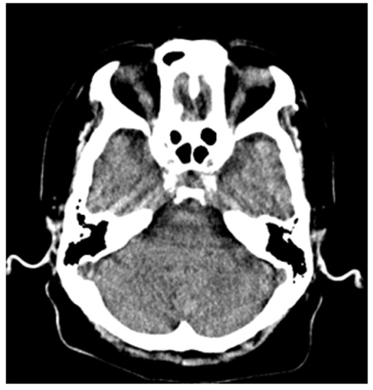
The present study reports a case of medullary metastasis without lung metastasis that occurred as a result of a malignant peripheral nerve sheath tumor (MPNST). An 81-year-old woman presented with a MPNST in the left brachial plexus, arising from the cervical nerve root. The patient underwent carbon ion radiotherapy; however, tumor recurrence was identified in the left shoulder. Subsequently, the patient underwent wide excision. Three weeks subsequent to surgery, imbalance and dysarthria developed suddenly. Dysphagia emerged and left upper limb pain disappeared on the day after symptom development. Magnetic resonance imaging (MRI) revealed that this was due to metastasis to the medulla. Five days subsequent to the onset of dysarthria, the patient succumbed due to respiratory failure. To the best of our knowledge, no previous cases of medullary metastasis arising from a MPNST in the absence of lung metastasis have been reported. MRI is a useful examination tool for the identification of brain metastases; however, the high cost of MRI as a routine examination must be considered due to the rarity of brain metastases. Therefore, methods to detect brain metastasis warrant further investigation.
Keywords: brainstem metastasis; malignant peripheral nerve sheath tumor.
Figures


References
-
- Valentin T, Le Cesne A, Ray-Coquard I, Italiano A, Decanter G, Bompas E, Isambert N, Thariat J, Linassier C, Bertucci F. Management and prognosis of malignant peripheral nerve sheath tumors: The experience of the French Sarcoma Group (GSF-GETO) Eur J Cancer. 2016;56:77–84. doi: 10.1016/j.ejca.2015.12.015. - DOI - PubMed
LinkOut - more resources
Full Text Sources
Other Literature Sources