Review
. 2016 Aug 31;4(1):57.
doi: 10.1186/s40560-016-0180-1. eCollection 2016.

Lung ultrasound-a primary survey of the acutely dyspneic patient

Affiliations
Review

Lung ultrasound-a primary survey of the acutely dyspneic patient

Francis Chun Yue Lee. J Intensive Care. .

Abstract

There has been an explosion of knowledge and application of clinical lung ultrasound (LUS) in the last decade. LUS has important applications in the ambulatory, emergency, and critical care settings and its deployability for immediate bedside assessment allows many acute lung conditions to be diagnosed and early interventional decisions made in a matter of minutes. This review detailed the scientific basis of LUS, the examination techniques, and summarises the current applications in several acute lung conditions. It is to be hoped that clinicians, after reviewing the evidence within this article, would see LUS as an important first-line modality in the primary evaluation of an acutely dyspneic patient.

Keywords: A-lines; B-lines; Curtain sign; I-lines; Lung ultrasound; Z-lines.


Figures
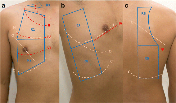
Fig. 1
Fig. 1
Scanning sectors (as used at the author’s centre). Zones on the right hemithorax. a R1 right anterior upper zone, R2 right anterior lower zone, Rs right supraclavicular fossa b R3 right lateral axilla zone, R4 right lateral lower zone c R5 right posterior upper zone R6 right posterior lower zone. I, II, III, IV first, second, third, fourth ribs, respectively, H horizontal fissure, O oblique fissure, C costophrenic recess, lowest limit of LUS study where curtain sign is found, *inferior angle of scapula
Fig. 2
Fig. 2
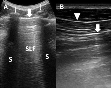
Comparing two scanning planes in LUS. a LUS performed in the longitudinal or cranio-caudal plane showed ribs (thin arrows) and their acoustic shadows (S). Just below the level of the ribs is the pleural line (thick arrow) and the sonographic lung field (SLF). b Subcutaneous tissue lines (arrowhead) could be mistaken for the pleural line (arrow) when LUS is performed in a transverse plane, without the guidance of the rib structure
Fig. 3
Fig. 3
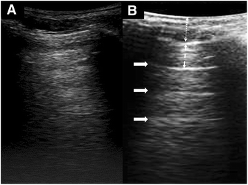
Two different appearances of air in LUS. a A hyperehoic appearance of lung air without A-lines. b LUS appearance with A-lines (solid arrows). The distance between the A-lines (dashed arrow) is equal to that between the transducer and the pleural line (dotted arrow). A-lines, other than that indicating a strong reflector is present, have no clinical significance
Fig. 4
Fig. 4
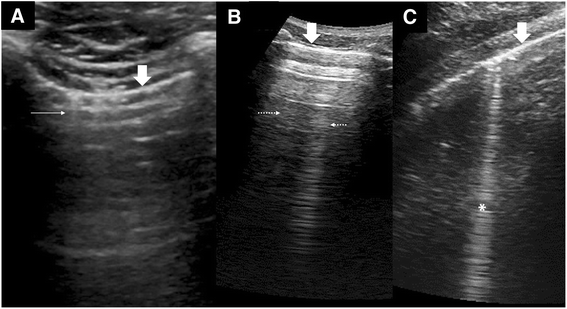
Vertical artifacts in LUS. a Lung comet (thin arrows) or I-lines arising from the pleural line (thick arrows) as seen with a high-frequency transducer at 8.5 MHz. b Static reverberation artifacts or Z-lines (dotted arrows) within the SLF are weak images with no relationship with the pleural line (thick arrow) and fades with depth. c A strong ring-down artifact or B-line (asterisk) starts from the pleural line (thick arrow) and reaches the depths without fading. It also swings side to side with lung sliding
Fig. 5
Fig. 5
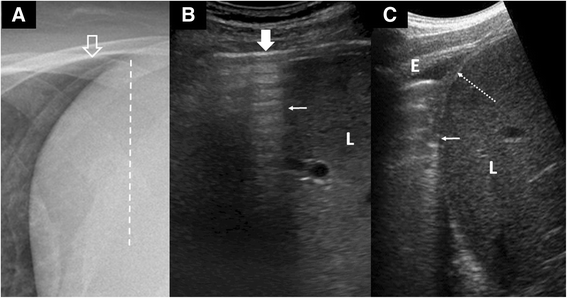
Curtain sign. a Chest X-ray illustrates the extent (dotted line) to which the lower parts of the lung (open arrow) cover the abdomen. b LUS shows the pleural line (solid arrow) ends abruptly with an edge (thin arrow) forming an acoustic shadow,  the “curtain sign,” which slides over the liver (L) with respiration. The lateral diaphragm is always hidden by the curtain and not seen in normal LUS. c An example of an abnormal curtain sign: a small effusion (E) causing an incomplete “curtain” sign (thin arrow) and exposing the lateral diaphragm (dotted arrow)
Fig. 6
Fig. 6
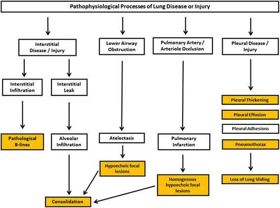
Pathological processes of lung disease and injury. This summarises some of the common endpoints of the pathological processes of lung disease and injury. The endpoints result in discernible features (yellow boxes) in LUS
Fig. 7
Fig. 7
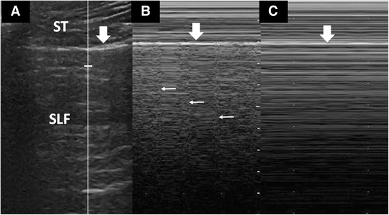
M-mode studies of lung sliding. a A proper M-mode study begins with the cursor (vertical line) centred over the SLF. The pleural line (thick arrow) separates the extra-pulmonary soft tissues (ST) and the SLF. b The M-mode showing “seashore” sign, where the quiet ST tracing (“sea”) is separated by the pleura line (thick arrow) from the noisy SLF tracing (“sandy shore”), caused by lung sliding. At regular intervals, the lung pulse (thin arrows) is seen. c M-mode showing “stratosphere” sign. The SLF tracing is “quiet” as there is no activity (lung sliding) at pleural line. There is also no lung pulse in this image
Fig. 8
Fig. 8
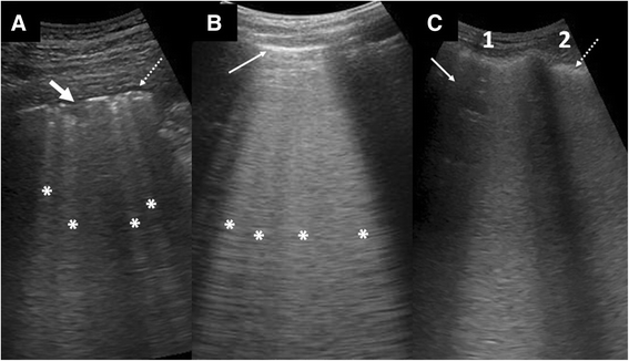
Examples of conditions with B-lines. a Pneumonia with several LUS features: B-lines (asterisk) of uneven spacing, a small consolidation (arrow), and small effusion (dotted arrow). b Cardiogenic pulmonary edema with many evenly spaced B-lines (asterisk) banded together into a thick sheet. Note the smooth and thin pleural line (thin arrow). c ARDS with dense B-lines involving two intercostal spaces (1, 2). Note that an area in 1 (arrow) is spared, indicating the patchy distribution of the disease process. The pleura is thickened and uneven (dotted arrow)
Fig. 9
Fig. 9
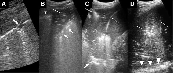
Features of consolidation. a Small consolidations appearing as subpleural defects (arrow). Ring-down artifacts or B-lines are also present (asterisks) b Wedge-shaped hypoechoic consolidations with trapped air within (thin arrow) and shred sign (thick arrow). A normal looking pleural line (open arrow head) and a thickened uneven pleural line (arrowhead) are shown. c A larger consolidation showing shred sign (thick arrow) and air bronchogram (thin arrow). Because this occurs at the lung base, the diaphragm (dotted arrow) is shown and hence the curtain sign is loss. d A lobar consolidation at the lung base showing air bronchogram (thin arrow), diaphragm (dotted arrow), and spine sign (arrowhead)
Fig. 10
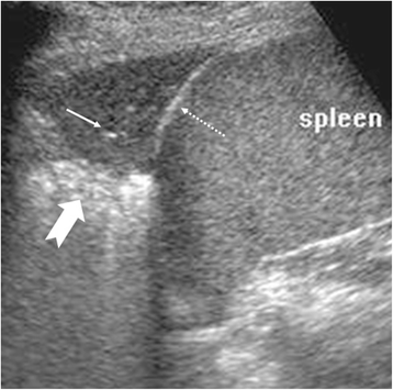
Fig. 10
Atelectasis. Hypoechoic homogenous lesion at the lung base with air bronchogram (thin arrow) and shred sign (thick arrow). The diaphragm (dotted arrow) is seen as the curtain sign is lost

References

    1. Lichtenstein D, Goldstein I, Mourgeon E, Cluzel P, Grenier P, Rouby JJ. Comparative diagnostic performances of auscultation, chest radiography and lung ultrasonography in acute respiratory distress syndrome. Anesthesiology. 2004;100:9–15. doi: 10.1097/00000542-200401000-00006. - DOI - PubMed
    1. Bourcier JE, Paquet J, Seinger M, Gallard E, Redonnet JP, Cheddadi F, et al. Performance comparison of lung ultrasound and chest x-ray for the diagnosis of pneumonia in the ED. Am J Emerg Med. 2014;32:115–8. doi: 10.1016/j.ajem.2013.10.003. - DOI - PubMed
    1. Pivetta E, Goffi A, Tizzani M, Porrino G, Ferreri E, Volpicelli G, et al. Lung ultrasound-implemented diagnosis of acute decompensated heart failure in the ED: A SIMEU multicentre study. Chest. 2015;148(1):202–10. doi: 10.1378/chest.14-2608. - DOI - PubMed
    1. Vopicelli G, Mussa A, Garofalo G, Cardinale L, Casoli G, Perotto F, et al. Bedside lung ultrasound in the assessment of alveolar-interstitial syndrome. Am J Emerg Med. 2006;24:689–96. doi: 10.1016/j.ajem.2006.02.013. - DOI - PubMed
    1. Milliner BHA, Tsung JW. An evidence-based lung ultrasound scanning protocol for diagnosing pediatric pneumonia. Ann Emerg Med. 2015;66(4):S107. doi: 10.1016/j.annemergmed.2015.07.330. - DOI

LinkOut - more resources