. 2016 Aug 18;9(8):1112-20.
doi: 10.18240/ijo.2016.08.05. eCollection 2016.

Inhibition of choroidal neovascularization by lentivirus-mediated PEDF gene transfer in rats

Affiliations

Inhibition of choroidal neovascularization by lentivirus-mediated PEDF gene transfer in rats

Ya-Jie Yu et al. Int J Ophthalmol. .

Abstract

Aim: To evaluate the effects of lentivirus-mediated pigment epithelium-derived factor (PEDF) gene transfer performed in treatment of rats with established choroidal neovascularization (CNV), and investigates the mechanism by which PEDF inhibits CNV in rats.

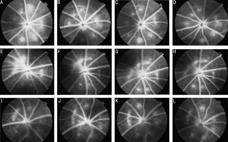
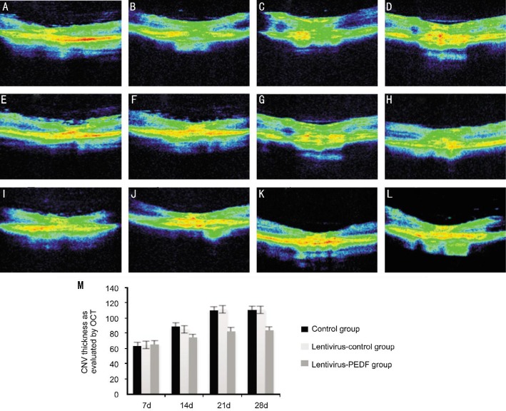
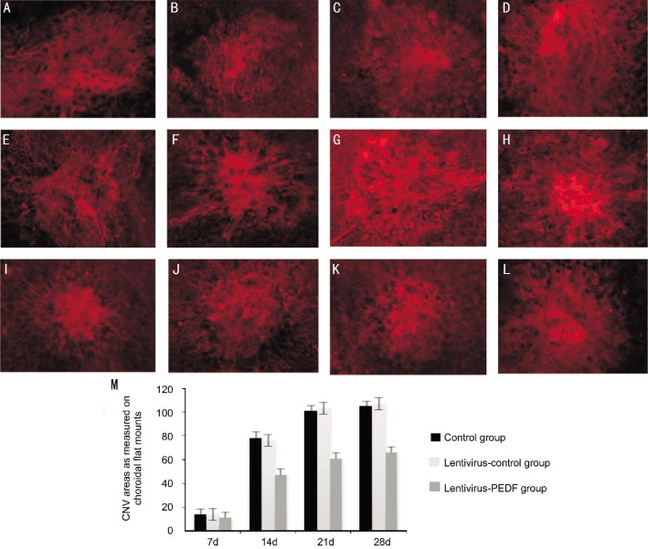
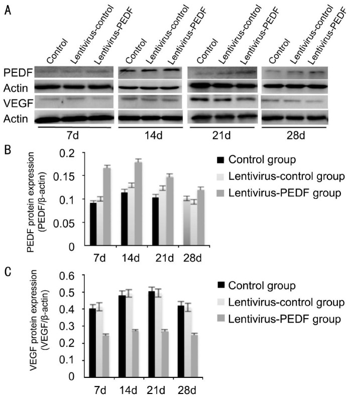
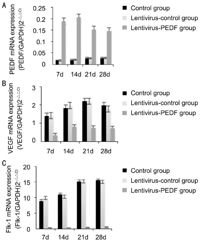
Methods: Brown Norway (BN) rats (n=204) were induced by exposure to a laser, and then randomly assigned to 3 groups: no treatment; treatments with intravitreal injection of lentivirus-PEDF-green fluorescent protein (GFP) or lentivirus-control GFP (free fluorescent protein). Following induction and treatment, the CNV tissue was assessed for form, size and vessel leakage by fluorescein fundus angiography (FFA), optical coherence tomography (OCT), histopathology, and examination of choroidal flat mounts. VEGF, Flk-1, and PEDF expression were evaluated by real-time polymerase chain reaction (PCR) and Western blot.

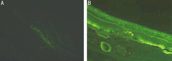
Results: A stable laser-induced rat model of CNV was successfully established, and used to demonstrate lentivirus-mediated PEDG gene transfer by intravitreal injection. Expression of green fluorescence labelled PEDF was observed in the retina up to 28d after injection. An intravitreal injection of lentivirus-PEDF-GFP at 7d led to a significant reduction in the size, thickness and area of CNV showed by FFA, OCT and choroidal flat mounts. PEDF was up-regulated while VEGF and Flk-1 were down-regulated in the lentivirus-PEDF-GFP group. The differences in VEGF and Flk-1 expression in the control and lentivirus-PEDF groups at 7, 14, 21 and 28d after laser induction were all statistically significant.

Conclusion: Lentivirus-mediated PEDF gene transfer is effective for use in treatment of laser-induced CNV, and PEDF exerts its therapeutic effects by inhibiting expression of VEGF and Flk-1.

Keywords: Flk-1; choroidal neovascularization; lentivirus; pigment epithelium-derived factor; vascular endothelial growth factor.


Figures

Figure 1
Figure 1. Ocular sections were examined by fluorescence microscopy on day 7 after injection
A: Representative image of fluorescence microscopy findings in the no treatment group; B: Representative image of fluorescence microscopy findings in the lentivirus-PEDF-GFP group. GFP fluorescence appeared in all retinal layers, and especially in photoreceptors and RPE cells.
Figure 2
Figure 2. FFA of CNV lesions
A-D: Representative late-phase FFA images of untreated laser-induced CNV areas at 7, 14, 21 and 28d after laser photocoagulation. FFA showed that laser-induced lesions partially resulted in emergence of CNV areas on day 7 after photocoagulation. From 14d to 28d, the CNV areas showed moderate-to-severe fluorescein leakage;E-H: Representative late-phase FFA images of control-lentivirus CNV areas at 7, 14, 21 and 28d after laser photocoagulation. The degree of angiographic leakage in this group was the same as that in the untreated laser-induced CNV group; I-L: Representative late-phase FFA images of CVN areas in rats which received lentivirus-PEDF. The images were taken at 7, 14, 21 and 28d after laser photocoagulation. FFA showed that an intravitreal injection of lentivirus-PEDF on day 7 after laser treatment led to a significant reduction in the size of the CNV area, as seen after fluorescein injection. Large and diffuse areas of leakage were not observed.
Figure 3
Figure 3. OCT images of CNV lesions
A-D: Representative OCT images of untreated laser-induced CNV taken at 7, 14, 21 and 28d after laser photocoagulation; E-H: Representative OCT images of control-lentivirus CNV taken at 7, 14, 21 and 28d after laser photocoagulation. The thickness and characteristics of the CNV in this group were the same as in the untreated laser-induced CNV group; I-L: Representative OCT images of lentivirus-PEDF CNV taken at 7, 14, 21 and 28d after laser photocoagulation. In this group, OCT showed significantly reduced thickness of the CNV; M: Comparison of CNV thickness in the three groups.
Figure 4
Figure 4. Choroidal flat mounts of CNV lesions
A-D: Representative images of choroidal flat mounts of untreated laser-induced CNV regions at 7, 14, 21 and 28d after laser photocoagulation; E-H: Representative images of choroidal flat mounts of control-lentivirus CNV at 7, 14, 21 and 28d after laser photocoagulation; I-L: Representative images of choroidal flat mounts of lentivirus-PEDF CNV at 7, 14, 21 and 28d after laser photocoagulation. The areas of the induced CNV lesions measured in choroidal flat mounts were smaller in eyes that received an intravitreal injection of lentivirus-PEDF on day 7 when compared to those areas in control eyes. All experiments were repeated 3 times, and data are expressed as the mean±SD (n=5 eyes). The differences between control and lentivirus-PEDF treated eyes remained statistically significant from day 7 to day 28 (P < 0.05).
Figure 5
Figure 5. Western blot analysis of PEDF and VEGF protein expression in the control group, lentivirus-control group, and lentivirus-PEDF group
A: The bands of PEDF and VEGF proteins in the control group, lentivirus-control group and lentivirus-PEDF group on days 7, 14, 21 and 28 after laser photocoagulation; B: Increased expression of PEDF in the lentivirus-PEDF group as compared with expression in the control group and lentivirus-control group from 7 to 28d after laser photocoagulation; C: Decreased VEGF expression in the lentivirus-PEDF group as compared with expression in the control group and lentivirus-control group from 7 to 28d after laser photocoagulation. All experiments were repeated 3 times.
Figure 6
Figure 6. Real-time PCR analysis of PEDF, VEGF and Flk-1 mRNA expression in the control-lentivirus group and lentivirus-PEDF group
A: When compared with the control group and lentivirus-control group, the level of PEDF mRNA expression in the lentivirus-PEDF-GFP group increased after day 7, and was markedly increased on day 14. A statistical analysis showed that PEDF expression in the lentivirus-PEDF-GFP group was significantly higher than that in the control group on days 7, 14, 21 and 28 (all P<0.05); B: Expression of VEGF mRNA in the lentivirus-PEDF group was decreased as compared with expression in the control group and lentivirus-control-GFP group from day 7 to day 28 after laser photocoagulation. The differences in VEGF expression between the control and lentivirus-PEDF-GFP groups on days 7, 14, 21 and 28 were all statistically significant (P<0.05); C: From day 7 to day 28 after laser photocoagulation, there was decreased expression of Flk-1 mRNA in the lentivirus-PEDF-GFP group as compared with expression in the control group and lentivirus-control-GFP group. The differences in Flk-1 mRNA expression between the control group and lentivirus-PEDF-GFP group on days 7, 14, 21 and 28 after photocoagulation were all statistically significant (P<0.05).

References

    1. Kawasaki R, Yasuda M, Song SJ, Chen SJ, Jonas JB, Wang JJ, Mitchell P, Wong TY. The prevalence of age-related macular degeneration in Asians: a systematic review and meta-analysis. Ophthalmology. 2010;117(5):921–927. - PubMed
    1. Kwak N, Okamoto B, Wood JM, Campochiaro PA. VEGF is major stimulator in modal of choroidal neovascularization. Invest Ophthalmol Vis Sci. 2000;41(10):3158–3164. - PubMed
    1. Dawson DW, Volpert OV, Gillis P, Crawford SE, Xu H, Benedict W, Bouck NP. Pigment epithelium-derived factor: a potent inhibitor of angiogenesis. Science. 1999;285(5425):245–248. - PubMed
    1. Renno RZ, Youssri AI, Michaud N, Gragoudas ES, Miller JW. Expression of Pigment Epithelium–Derived Factor in Experimental Choroidal Neovascularization. Invest Ophthalmol Vis Sci. 2002;43(5):1574–1580. - PubMed
    1. Chen G, Li W, Tzekov R, Mao S, Tong Y. Bevacizumab versus ranibizumab for neovascular age-related macular degeneration: a meta-analysis of randomized controlled trials. Retina. 2015;35(2):187–193. - PubMed

LinkOut - more resources