. 2016 Aug 18;1(13):e88782.
doi: 10.1172/jci.insight.88782.

Redefined clinical features and diagnostic criteria in autoimmune polyendocrinopathy-candidiasis-ectodermal dystrophy

Affiliations

Redefined clinical features and diagnostic criteria in autoimmune polyendocrinopathy-candidiasis-ectodermal dystrophy

Elise M N Ferre et al. JCI Insight. .

Abstract

Autoimmune polyendocrinopathy-candidiasis-ectodermal dystrophy (APECED) is a rare primary immunodeficiency disorder typically caused by homozygous AIRE mutations. It classically presents with chronic mucocutaneous candidiasis and autoimmunity that primarily targets endocrine tissues; hypoparathyroidism and adrenal insufficiency are most common. Developing any two of these classic triad manifestations establishes the diagnosis. Although widely recognized in Europe, where nonendocrine autoimmune manifestations are uncommon, APECED is less defined in patients from the Western Hemisphere. We enrolled 35 consecutive American APECED patients (33 from the US) in a prospective observational natural history study and systematically examined their genetic, clinical, autoantibody, and immunological characteristics. Most patients were compound heterozygous; the most common AIRE mutation was c.967_979del13. All but one patient had anti-IFN-ω autoantibodies, including 4 of 5 patients without biallelic AIRE mutations. Urticarial eruption, hepatitis, gastritis, intestinal dysfunction, pneumonitis, and Sjögren's-like syndrome, uncommon entities in European APECED cohorts, affected 40%-80% of American cases. Development of a classic diagnostic dyad was delayed at mean 7.38 years. Eighty percent of patients developed a median of 3 non-triad manifestations before a diagnostic dyad. Only 20% of patients had their first two manifestations among the classic triad. Urticarial eruption, intestinal dysfunction, and enamel hypoplasia were prominent among early manifestations. Patients exhibited expanded peripheral CD4+ T cells and CD21loCD38lo B lymphocytes. In summary, American APECED patients develop a diverse syndrome, with dramatic enrichment in organ-specific nonendocrine manifestations starting early in life, compared with European patients. Incorporation of these new manifestations into American diagnostic criteria would accelerate diagnosis by approximately 4 years and potentially prevent life-threatening endocrine complications.


Conflict of interest statement

The authors have declared that no conflict of interest exists.

Figures

Figure 1
Figure 1. Enrichment of nonendocrine autoimmune manifestations in American autoimmune polyendocrinopathy-candidiasis-ectodermal dystrophy (APECED).
(A) Prevalence of all disease manifestations in the 35 American APECED patients. The black bars represent the prominent hexad of nonendocrine organ-specific manifestations that is enriched in our cohort relative to all other reported experiences. (BD) Distribution of all clinical manifestations (B), endocrinopathies (C), and nonendocrine manifestations (D) among the 35 American APECED patients. (E) Representative image from a liver biopsy of an APECED patient with autoimmune hepatitis showing severe chronic hepatitis with expansion of portal areas by inflammation and fibrosis. There is extensive interface hepatitis with numerous plasma cells infiltrating into the hepatic parenchyma (H&E; scale bar: 50 μm; original magnification, ×200). (F) Representative image from a stomach biopsy of an APECED patient with autoimmune gastritis showing chronic antral inflammation with lymphoplasmacytic infiltrates in the lamina propria and occasionally on glands (H&E, scale bar: 50 μm; original magnification, ×200). (G) Representative image from an open lung biopsy of an APECED patient with autoimmune pneumonitis showing chronic bronchiolitis with lymphocytic infiltration within and around the bronchiolar mucosa and lymphoid aggregates in the interstitium nearby (H&E, scale bar: 50 μm; original magnification, ×200). (H) Representative image from a minor salivary gland biopsy in an APECED patient with Sjögren’s-like syndrome showing lymphocytic and plasma cell infiltration in and around the ducts of the gland (H&E; scale bar: 50 μm; original magnification, ×200).
Figure 2
Figure 2. Early manifestations and redefined diagnostic criteria in American autoimmune polyendocrinopathy-candidiasis-ectodermal dystrophy (APECED).
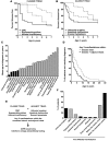
(A and B) Prevalence by age of the classic triad manifestations (A) and the adjunct triad of urticarial eruption, intestinal dysfunction, and enamel hypoplasia (B) within the first 7 years of life, before the mean age at which a diagnostic dyad was reached in the 35 American APECED patients. (C) Mean age at diagnosis of all clinical manifestations among the APECED patients who developed the corresponding disease components. Black bars denote the clinical manifestations with a mean age of diagnosis within the first 7 years of life, before the mean age at which a classic diagnostic dyad is reached. (D) Age of reaching diagnosis based on development of any 2 classic triad manifestations (current diagnostic criteria) versus reaching the diagnosis based on development of any 2 manifestations within the combined classic triad and adjunct triad of urticarial eruption, intestinal dysfunction, and enamel hypoplasia (expanded diagnostic criteria). (E) Redefined expanded diagnostic criteria and diagnostic algorithm aimed at promoting earlier diagnosis of American APECED patients. (F) Distribution of the initial manifestation in the 35 American APECED patients. Gray bars denote manifestations within the current classic diagnostic triad.
Figure 3
Figure 3. Clinical and histological presentation of urticarial eruption in American children with autoimmune polyendocrinopathy-candidiasis-ectodermal dystrophy (APECED).
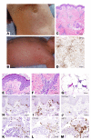
(A and B) Representative images showing erythematous urticarial eruption on the torsos of two children with APECED. (C) Histologic examination of skin biopsies reveals perivascular and periadnexal inflammation in the superficial and deep dermis with pallor of the papillary dermis (H&E; scale bar: 2 mm; original magnification, ×40) (D) Myeloperoxidase (MPO) immunohistochemical staining highlights the presence of neutrophils and karyorrhectic debris derived from neutrophils (scale bar: 2 mm; original magnification, ×40). (E) High-magnification view of the epidermis and superficial dermis revealing focal interface vacuolar changes along the epidermal basement membrane zone, with thickened and irregular basement membrane and superficial perivascular inflammation, with karyorrhectic debris in the dermis (H&E; scale bar: 200 μm; original magnification, ×200). (F) High-magnification view of the reticular dermis revealing perivascular and interstitial inflammation predominantly composed of mononuclear cells with karyorrhectic debris, while mature neutrophils are infrequent (H&E; scale bar: 200 μm; original magnification, ×200). (G) High-magnification view of the subcutaneous adipose tissue revealing inflammation predominantly composed of mononuclear cells with karyorrhectic debris (H&E; scale bar: 300 μm; original magnification, ×200). (HJ) Immunohistochemical staining with lymphocyte markers (CD3, H; CD8, I; CD79a, J; CD4, not shown) revealing lymphocytic exocytosis with CD4+ and CD8+ T lymphocytes lining up along the base of epidermis and occasional cells within the spinous layers; CD79a+ B lymphocytes are rarely seen in a perivascular distribution (scale bar: 200 μm; original magnification, ×400). (KM) Immunohistochemical staining with lymphocyte markers (CD3, K), macrophage/monocyte/histiocyte markers (CD163, L; CD68, not shown), and neutrophil/myeloid lineage markers (MPO, M) revealing periadnexal/perieccrine inflammation composed of abundant neutrophils/myeloid cells with karyorrhexis and scattered lymphocytes and monocytes/macrophages/histiocytes (scale bar: 200 μm; original magnification, ×400).
Figure 4
Figure 4. Expansion of CD21loCD38lo B cells in the peripheral blood of autoimmune polyendocrinopathy-candidiasis-ectodermal dystrophy (APECED) patients and Aire–/– mice.
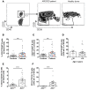
(A) Gating strategy and representative FACS plots of CD21loCD38lo B cells in peripheral blood. After initial FSC/SCC gating of peripheral blood mononuclear cells (not shown), CD19+ B cells were selected (left) and CD21loCD38lo B cells were delineated and quantified. Representative FACS plots of CD21loCD38lo B cells from an APECED patient (middle) and a healthy donor (right) showing enrichment of these cells in patient peripheral blood. (B and C) Summary data for the percentage of CD21loCD38lo B cells within total CD19+ B cells (B, ***P = 0.0004, Mann-Whitney test) and for the absolute number of CD21loCD38lo B cells per μl of human blood (C, ***P = 0.0007, Mann-Whitney test). APECED patients, n = 30; healthy donors, n = 40. The 23 gray squares correspond to the results obtained in the 23 APECED patients with biallelic AIRE mutations that include c.967_979del13 in heterozygosity or homozygosity (group A), the 3 blue squares correspond to the results obtained in the 3 APECED patients with biallelic AIRE mutations other than c.967_979del13 (group B), and the 4 red squares correspond to the results obtained in the 4 APECED patients without biallelic AIRE mutations (group C). The percentage and absolute number of CD21loCD38lo B cells did not significantly differ between patient groups A, B, and C (unpaired t test). (D) Summary data for the absolute number of CD21loCD38lo B cells per μl of blood of APECED patients that are younger (n = 5) or older (n = 25) than 10 years. (E and F) Summary data for the percentage of CD11c+CD11b+ B cells within total CD19+ B cells (E, ****P < 0.0001, Mann-Whitney test) and for the absolute number of CD11c+CD11b+ B cells per μl of mouse blood (F, ****P < 0.0001, Mann-Whitney test). Aire+/+ mice, n = 16; Aire–/– mice, n = 17; 4 independent experiments. All quantitative data represent mean ± SEM.

References

    1. Anderson MS, et al. Projection of an immunological self shadow within the thymus by the aire protein. Science. 2002;298(5597):1395–1401. doi: 10.1126/science.1075958. - DOI - PubMed
    1. Mathis D, Benoist C. Aire. Annu Rev Immunol. 2009;27:287–312. doi: 10.1146/annurev.immunol.25.022106.141532. - DOI - PubMed
    1. Oftedal BE, et al. Dominant mutations in the autoimmune regulator AIRE are associated with common organ-specific autoimmune diseases. Immunity. 2015;42(6):1185–1196. doi: 10.1016/j.immuni.2015.04.021. - DOI - PubMed
    1. Cetani F, et al. A novel mutation of the autoimmune regulator gene in an Italian kindred with autoimmune polyendocrinopathy-candidiasis-ectodermal dystrophy, acting in a dominant fashion and strongly cosegregating with hypothyroid autoimmune thyroiditis. J Clin Endocrinol Metab. 2001;86(10):4747–4752. doi: 10.1210/jcem.86.10.7884. - DOI - PubMed
    1. Su MA, et al. Mechanisms of an autoimmunity syndrome in mice caused by a dominant mutation in Aire. J Clin Invest. 2008;118(5):1712–1726. doi: 10.1172/JCI34523. - DOI - PMC - PubMed