

Involvement of let-7 microRNA for the therapeutic effects of Rhenium-188-embedded liposomal nanoparticles on orthotopic human head and neck cancer model
- PMID: 27588466
- PMCID: PMC5323192
- DOI: 10.18632/oncotarget.11666
Involvement of let-7 microRNA for the therapeutic effects of Rhenium-188-embedded liposomal nanoparticles on orthotopic human head and neck cancer model
Abstract
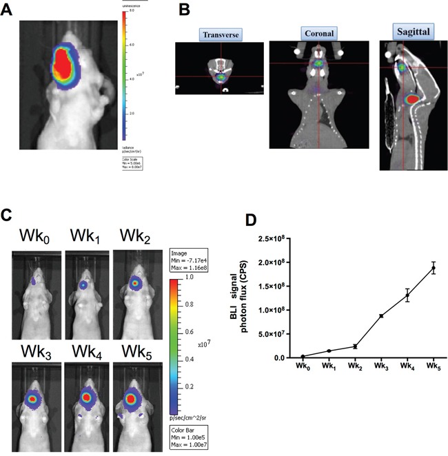
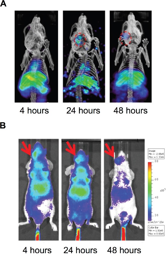
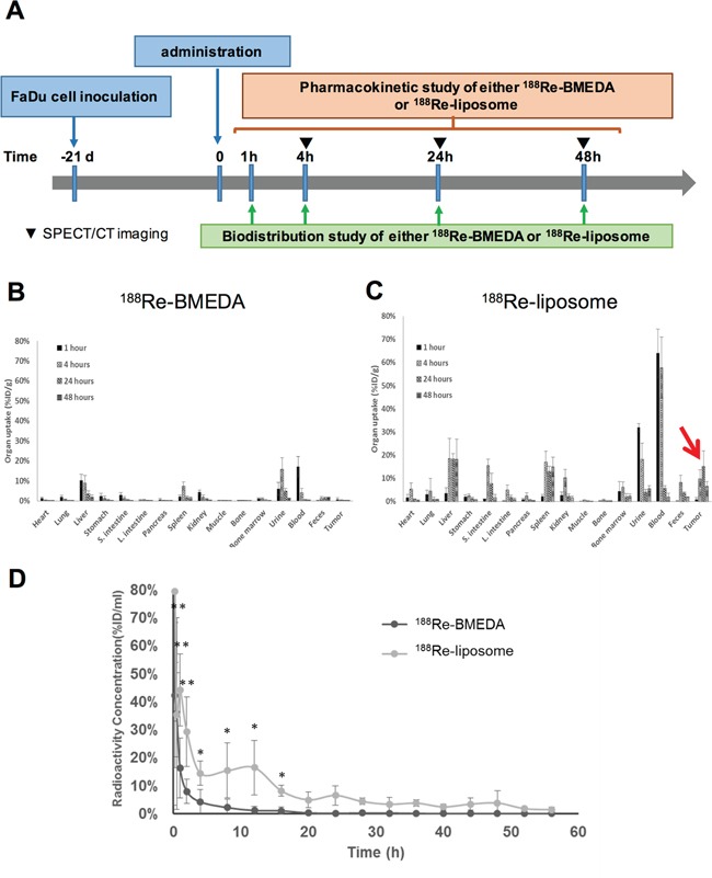
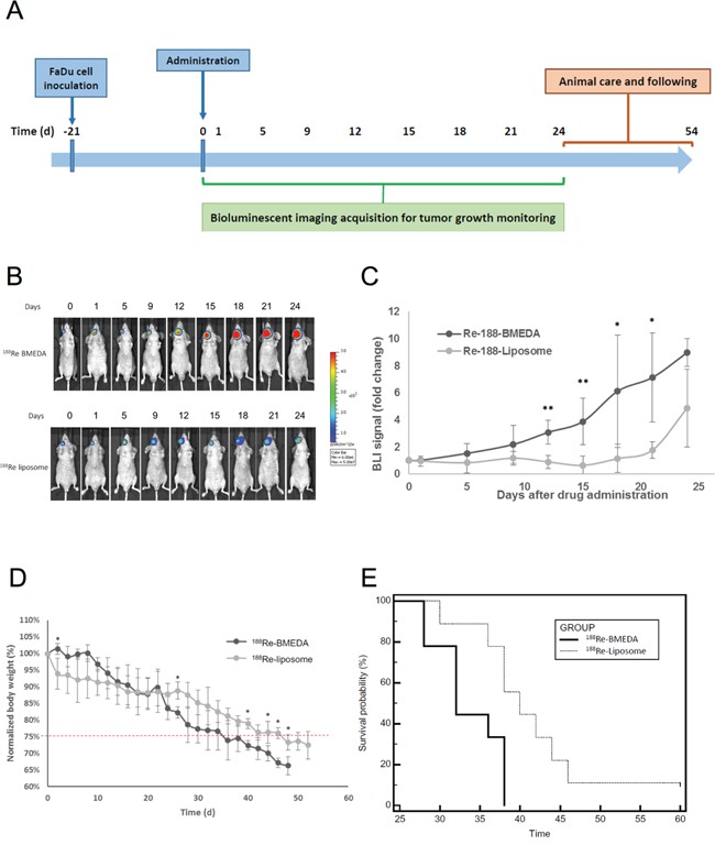
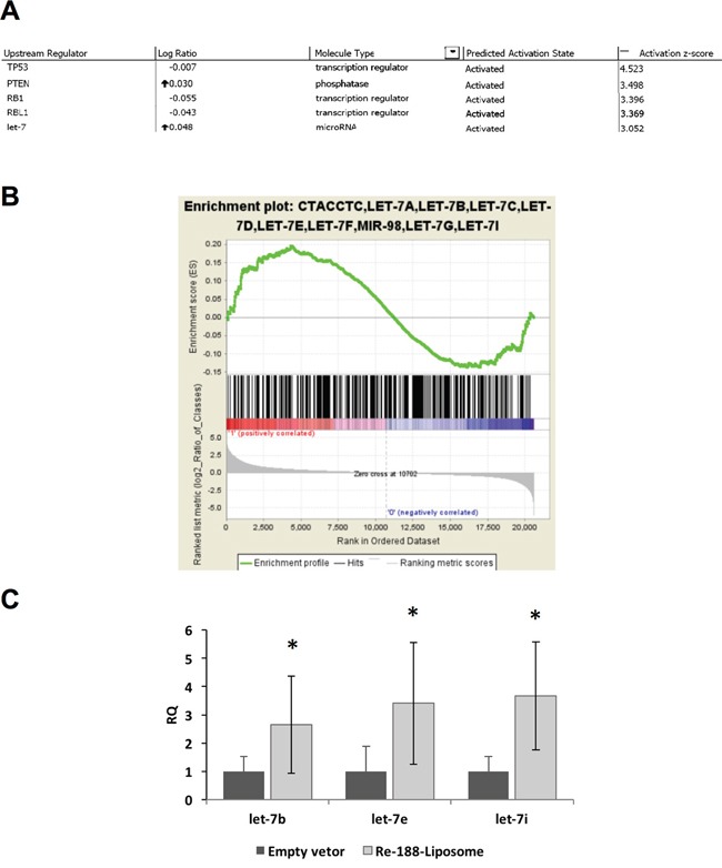
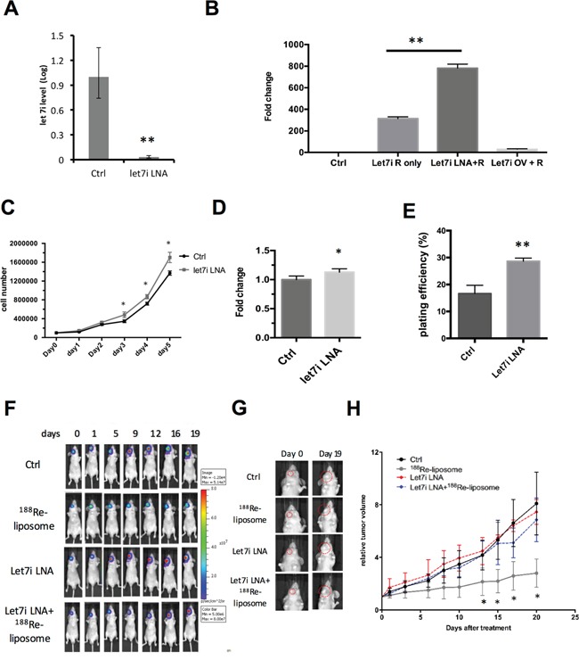
Human head and neck squamous cell carcinoma (HNSCC) is usually treated by surgical resection with adjuvant radio-chemotherapy. In this study, we examined whether the radiopharmaceutical 188Re-liposome could suppress the growth of HNSCC followed by an investigation of molecular mechanisms. The orthotopic HNSCC tumor model was established by human hypopharyngeal FaDu carcinoma cells harboring multiple reporter genes. The drug targeting and therapeutic efficacy of 188Re-liposome were examined using in vivo imaging, bio-distribution, pharmacokinetics, and dosimetry. The results showed that 188Re-liposome significantly accumulated in the tumor lesion compared to free 188Re. The circulation time and tumor targeting of 188Re-liposome were also longer than that of free 188Re in tumor-bearing mice. The tumor growth was suppressed by 188Re-liposome up to three weeks using a single dose treatment. Subsequently, microarray analysis followed by Ingenuity Pathway Analysis (IPA) showed that tumor suppressor let-7 microRNA could be an upstream regulator induced by 188Re-liposome to regulate downstream genes. Additionally, inhibition of let-7i could reduce the effects of 188Re-liposome on suppression of tumor growth, suggesting that let-7 family was involved in 188Re-liposome mediated suppression of tumor growth in vivo. Our data suggest that 188Re-liposome could be a novel strategy for targeting HNSCC partially via induction of let-7 microRNA.
Keywords: 188Re-liposome; HNSCC; let-7 microRNA; microarray analysis; orthotopic tumor model.
Conflict of interest statement
No other potential conflicts of interest relevant to this article was reported.
Figures
Similar articles
-
Evaluation of 188Re-labeled PEGylated nanoliposome as a radionuclide therapeutic agent in an orthotopic glioma-bearing rat model.Int J Nanomedicine. 2015 Jan 9;10:463-73. doi: 10.2147/IJN.S75955. eCollection 2015. Int J Nanomedicine. 2015. PMID: 25624760 Free PMC article.
-
Involvement of Differentially Expressed microRNAs in the PEGylated Liposome Encapsulated 188Rhenium-Mediated Suppression of Orthotopic Hypopharyngeal Tumor.Molecules. 2020 Aug 8;25(16):3609. doi: 10.3390/molecules25163609. Molecules. 2020. PMID: 32784458 Free PMC article.
-
Molecular imaging and therapeutic efficacy of 188Re-(DXR)-liposome-BBN in AR42J pancreatic tumor-bearing mice.Oncol Rep. 2012 Nov;28(5):1736-42. doi: 10.3892/or.2012.1978. Epub 2012 Aug 22. Oncol Rep. 2012. PMID: 22922965
-
Translating Research for the Radiotheranostics of Nanotargeted 188Re-Liposome.Int J Mol Sci. 2021 Apr 8;22(8):3868. doi: 10.3390/ijms22083868. Int J Mol Sci. 2021. PMID: 33918011 Free PMC article. Review.
-
Absorbed doses in humans from 188Re-Rituximab in the free form and bound to superparamagnetic iron oxide nanoparticles: Biodistribution study in mice.Appl Radiat Isot. 2018 Jan;131:96-102. doi: 10.1016/j.apradiso.2017.10.041. Epub 2017 Oct 23. Appl Radiat Isot. 2018. PMID: 29173814 Review.
Cited by
-
Characterization and profiling of the microRNA in small extracellular vesicles isolated from goat milk samples collected during the first week postpartum.JDS Commun. 2023 Aug 19;4(6):507-512. doi: 10.3168/jdsc.2022-0369. eCollection 2023 Nov. JDS Commun. 2023. PMID: 38045901 Free PMC article.
-
PEGylated liposome-encapsulated rhenium-188 radiopharmaceutical inhibits proliferation and epithelial-mesenchymal transition of human head and neck cancer cells in vivo with repeated therapy.Cell Death Discov. 2018 Oct 31;4:100. doi: 10.1038/s41420-018-0116-8. eCollection 2018. Cell Death Discov. 2018. PMID: 30393570 Free PMC article.
-
Brachytherapy Approach Using 177Lu Conjugated Gold Nanostars and Evaluation of Biodistribution, Tumor Retention, Dosimetry and Therapeutic Efficacy in Head and Neck Tumor Model.Pharmaceutics. 2021 Nov 9;13(11):1903. doi: 10.3390/pharmaceutics13111903. Pharmaceutics. 2021. PMID: 34834318 Free PMC article.
-
Adenovirotherapy Delivering Cytokine and Checkpoint Inhibitor Augments CAR T Cells against Metastatic Head and Neck Cancer.Mol Ther. 2017 Nov 1;25(11):2440-2451. doi: 10.1016/j.ymthe.2017.09.010. Epub 2017 Sep 14. Mol Ther. 2017. PMID: 28974431 Free PMC article.
-
Insights into Nanomedicine for Head and Neck Cancer Diagnosis and Treatment.Materials (Basel). 2022 Mar 11;15(6):2086. doi: 10.3390/ma15062086. Materials (Basel). 2022. PMID: 35329542 Free PMC article. Review.
References
-
- Bray F, Jemal A, Grey N, Ferlay J, Forman D. Global cancer transitions according to the Human Development Index (2008-2030): a population-based study. The lancet oncology. 2012;13:790–801. - PubMed
-
- Casciato DA, Territo MC. A Lippincott manual. Philadelphia: Wolters Kluwer Health/Lippincott Williams & Wilkins; 2012. Manual of clinical oncology; p. 1. online resource (x 928 p)
-
- Nestor MV. Targeted radionuclide therapy in head and neck cancer. Head Neck. 2010;32:666–678. - PubMed
-
- Samad A, Sultana Y, Aqil M. Liposomal drug delivery systems: an update review. Curr Drug Deliv. 2007;4:297–305. - PubMed
-
- Milla P, Dosio F, Cattel L. PEGylation of proteins and liposomes: a powerful and flexible strategy to improve the drug delivery. Curr Drug Metab. 2012;13:105–119. - PubMed
MeSH terms
Substances
LinkOut - more resources
Full Text Sources
Other Literature Sources
Medical
